Prevalence of Azo Dye Adulterated Palm Oil in Ondo State (Nigeria) and Toxicological Effects on Liver, Kidney and Testicular Tissues of Albino Rats
Kola-Ajibade Ibukun R1*, Olusola Augustine O2
Department of Biochemistry, Faculty of Science, Adekunle Ajasin University Akungba Akoko Ondo State, Nigeria.
- *Corresponding Author:
- Kola-Ajibade Ibukun
Department of Biochemistry, Faculty of Science, Adekunle Ajasin University Akungba Akoko Ondo State, Nigeria.
Tel: 07032738869
E-mail: ibukunkolaajibade@gmail.com
Received Date: December 03, 2021; Accepted Date: December 16, 2021; Published Date: December 23, 2021
Citation: Ibukun AKR, Augustine OO (2021) Prevalence of Azo Dye Adulterated Palm Oil in Ondo State (Nigeria) and ToxicologiEcffale cts on Liver, Kidney and Testicular Tissues of Albino Rats. JFST Vol:5 No:4
Abstract
Adulteration of fats and oils is an ancient challenge and has been the subject of many studies. Due to the recent increase in consumption of palm oil, its demand is outweighing consistent supply in Nigeria, adulteration has become imminent. The widening gap has given way to adulteration with azo dyes especially with Sudan III and IV dyes. This study was undertaken to determine the prevalence of adulteration of palm oil with azo dyes across the senatorial districts of Ondo State, Southwest Nigeria, and to evaluate its toxicity on the liver, kidney, and testicular tissues in exposed male albino rats. Palm oil sourced from oil mills and open markets across the senatorial districts were analyzed for the presence of adulterants. Twenty-five albino rats were divided into five groups and treated as thus; group I (control), groups II and III (1 ml/kg of unadulterated and adulterated palm oil respectively), groups IV and V (50 mg/kg Sudan III and IV respectively) for 28 days. At the end of the experiment, animals were sacrificed; kidney, liver and testis tissues were extracted for histological examination. The result showed that none of the palm oil samples from Oil mills contained adulterants, while some of the samples from open markets had adulterants. The source of adulteration was mainly from bulk buyers and retailers which is also more prevalent in the urban areas of the State. Histological examination showed micro morphological alteration characterized by mild to severe cellular damage and the presence of red inflammatory cells in the tissues. It is recommended that consumers patronize palm oil sellers at oil mills which is the genuine source and the government of Ondo State should create a public health awareness to consumers on the deleterious effects of consumption.
Keywords
Azo dye; Palm oil; Prevalence; Adulteration; Histological examination
Introduction
Oil palm (Elaeis guineensis) is a monocotyledonous plant belonging to the palm family Arecaceae, it produces male and female inflorescences in an alternating cycle [1]. The fruit produces two distinct types of oil: orange-red crude palm oil which is extracted from the mesocarp and brownish yellow crude palm kernel oil extracted from the seeds (kernel). The oil palm plant has the highest oil producing capacity with an average 3.5 tons and its demand is on the increase in West Africa [2]. Palm oil is produced in forty- two (42) countries globally and Nigeria is the fourth largest producer [3,4]. The oil palm (Elaeis guinensis) is generally believed to have originated from the rainforest region of West Africa [5] with the highest allelic diversity found in Nigerian and they were genetically similar to those found in Bahia Brazil, indicating that Nigeria is the possible center of origin of oil palm [6]. Palm oil manufacturing represents one of the most effective methods of alleviating Nigerians from poverty and ensuring food security. It has provided employment for millions of unskilled and semi-skilled people.
Food Adulteration is a process of reducing the quality of food by intentional or unintentional substitution of food with some inferior foreign substance or by removal of some value added food substitute from main food item [7]. Food fraud is currently a persistent global problem with improved technology and no food substance is exempted as in the case of palm oil adulteration [8]. Adulteration of fats and oils is an old problem and has been the subject of many studies [9,10]. The "get rich quick syndrome’ is a canker worn that has eaten deep in Nigerians, making ‘us’ to forget the right procedure of doing things and thus high inflow of adulterated health related consumables in the Nigerian market stressing on its socio economic consequence to the society at large. Moreover, the perpetrators of this crime are exploiting the glaring lapses in the regulatory supervision by the agency statutorily charged with keeping an eye on the quality of foods consumed in the country. Nigerians are therefore, left at the mercy of dubious dealers in substandard and adulterated food items, with obvious long-term health implications for the consumers. In recent years, the increasing demand for palm oil in Nigeria has out - weighed supply leading to a widening gap between demand and production and this has led to inevitable increase in the level of adulteration [11,12].
It has been reported that azo dyes (Sudan III and IV) are been added to palm oil to improve the color and make it more appealing to consumers [8,12,13]. Sudan dyes are considered illegal dyes labelled group 3 carcinogens and their illegal addition to palm oil has been reported [13-15].
It has been observed that most edible palm oil in Nigeria fall short of the recommended quality standards that is considered safe for consumption [12,16]. The low quality could be as a result of the presence of some inclusion which has been added intentionally by the producers or marketers to enhance quantity, appearance and viscosity. In West Africa, the manufacturing and processing of palm oil is done on small, medium and large scale, it is therefore almost impossible to detect fraud in the system [8,17]. However, a major disadvantage associated with the use of adulterants in palm oil is that the adulterants have not undergone adequate research and the degree of health hazards they can pose to humans when consumed not well established [18,19].
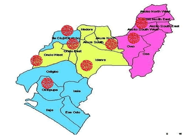
This study however aims to determine the prevalence and source (point) of adulterated palm oil across the senatorial districts in Ondo state Nigeria and histological examination for probable toxicological effects on the kidney, liver and testicular tissues (Figure I- samples sourced from areas marked in red).
Materials and methodology
Samples
palm oil, Sudan III, Sudan IV, albino rats
Reagents
Tween 20, petroleum ether, Hydrochloric acid, formalin
Test for adulteration of palm oil.
Forty (40) samples of palm oil were sourced from oil mills and open markets (areas marked in red from Figure I) twenty- six (26) samples from open markets and fourteen (14) from Oil mils (factories) across Ondo State. The samples were left to stand on the bench for six (6) months in transparent pet bottles by the end of 6 months; red dyes were seen settled at the base of palm oil bottles containing adulterants.
Chemical test – the method used by Nwachoko and Fortune, [20] was also employed to confirm the presence of adulterant – Sudan dye in samples of palm oil.This analysis was done using petroleum spirit and concentrations of hydrochloric acid/water (1:1). To 5 ml of each oil sample in different test tubes, 15 ml of petroleum ether was added followed by the 5 ml hydrochloric acid to different test tubes. Different shades of yellow were observed at the top and a clear /colorless base indicating the absence of adulterants while samples containing adulterants showed a reddish yellow top and a reddish clear base.
Dye sample preparation
Sudan dyes are not generally soluble in distilled water. Total solubility was achieved by dissolution in 10% Tween 20. The solution obtained was administered orally to the rats using an intra-gastric feeding syringe.
Experimental grouping/treatment of animals–25 male albino rats were weighing 111 g- 198 g were assigned into 5 groups (I,II,III,IV, and V). Five animals in each cage, they were acclimatized to their environment and diet for 7 days. Groups II, III, IV and V were the test groups. Group I was the control group. Groups II and III were given 1 ml/kg of unadulterated palm oil and adulterated respectively [20]. Groups IV and V were given 50 mg/kg Sudan III and Sudan IV respectively [21].
Histological examination method
The kidney, Liver and Testes of the control and treated groups were immediately extracted and put in formalin and Bouin’s fliud (Testes) respectively. The sections were stained using haematoxylin and eosin method.
Results
| Senatorial location | Adulterated samples found | unadulterated samples found | Total number of sample (n) |
|---|---|---|---|
| Central(Akure, Ogbese, Ondo, Idanre) | 7 | 8 | 15 |
| South(Okitipupa , Oke igbo) | 1 | 10 | 11 |
| North(Akoko- Supare, Akungba, Ayegunle – Oba, Arigidi, Emure, Owo) | 4 | 10 | 14 |
Table 1: Classification based on senatorial districts.
| Source | Number of Adulterated samples | Number of unadulterated samples | Total number of samples found (n) |
|---|---|---|---|
| Factory | None | 14 | 14 |
| Open market | 12 | 14 | 26 |
Table 2: Classification based on source (point) of collection.
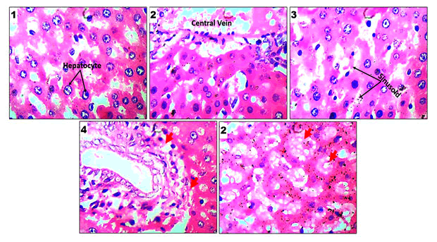
Show normal central venules without congestion, the morphology of the hepatocytes appears normal with distinct layering, the sinusoids appear normal and not infiltrated, portal triad appear intact. Groups with significant observable alteration is marked with red arrows and are characterized by infiltrated and congested sinusoids (mild in 3 and becomes conspicuous in 4 and 5), portal dilation, severe hemorrhage especially in groups 4 and 5 , steatosis (4 and 5), portal inflammation, reduced hepatocytes, seen in 5 are pyknotic hepatocytes surrounded by white spaces and some signs of fibrosis across 3-5 groups.
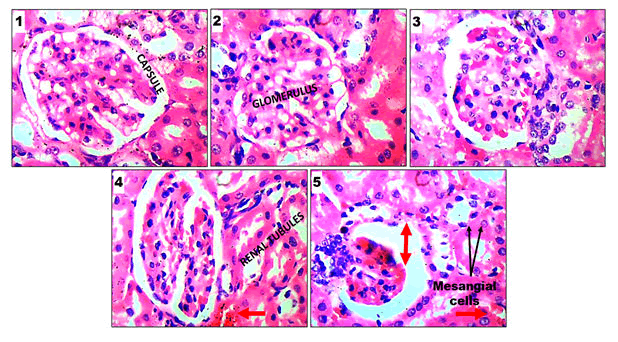
Group 1 (CONTROL), Group 2 (unadulterated) GRP 3 (adulterated), Group 4 (Sudan III), Group 5 (Sudan IV). Groups 1 and 2 show normal architecture as seen: the renal cortex shows normal glomeruli, mesangial cells and capsular spaces, the renal tubules and, the interstitial spaces appear normal with no observable significant dilation, necrosis or hemorrhage. Groups marked with red arrows indicates micro morphological alteration characterized by mild to severe cellular loos, pyknotic mesangial cells, presence of red inflammatory cells, mild to severe hemorrhage, glomerulosclerosis, wider capsular space as well as collapsed renal tubules.
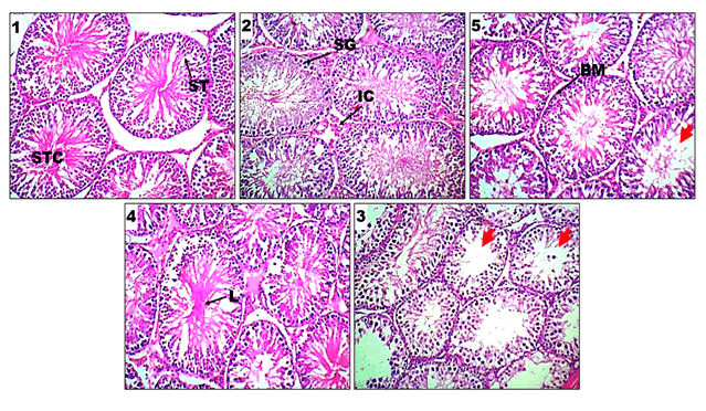
The seminiferous tubules ST, spermatogonium SG (containing primary, secondary and tertiary spermatocytes), basement membrane BM, interstitial cells IC, lumen L, spermatocytes STC, are all visible across the various groups. Seminiferous tubules appear intact with full lumen as well as defined Sertoli cells seen within the lumen in groups 1 & 2. Groups 3, 4 and 5 showed mild to severe micromorphological alterations characterized with observable interstitial space distortion (widen space), pyknotic interstitial cells; which is indication for loss of Leydig cells, Incomplete progression of spermatogonia within the seminiferous tubules (5): indicating spermatogenic arrest (incomplete maturation of sperm cells) as seen in the lumen characterized with empty content, degenerating spermatogia cell nuclei as well as scanty luminal content. Basement membrane appear to be mildly diminished. Seminiferous tubules appear empty with scanty Sertoli cells.
Discussion
Result from (Table I) shows that from a total of forty (40) samples of palm oil collected across the three senatorial districts of Ondo state, twelve (12) were found to contain adulterants with the highest prevalence found in Ondo Central. Adulteration of palm oil with Sudan dyes has been reported by many studies and a wide speculation that crude palm oil is being adulterated in Nigeria [8, 12,19,21,22].
This is the first study to check for prevalence of adulterated palm oil in Ondo state, we were able to establish the highest prevalence of azo dye adulterated palm oil in Ondo central (specifically Akure). The probable reason for higher prevalence in Ondo central is because it is a more commercialized area (urban area) with higher population, leading to demand out weighing supply. Therefore, retailers in a bid to meet up with the rising demand, they venture into adulteration. This is in agreement with [10] who observed that the consumption of palm oil (demand) is growing faster than its supply and widening gap between demand and supply has been accompanied by increasing reports of adulteration.
Moreover, results obtained from (Table II), none of the fourteen (14) samples collected from Oil mills (factories) contained Sudan dyes. We were able to rule out Oil mills as a possible source or point of palm oil adulteration. It has become obvious that the adulteration is majorly practiced by bulk buyers and retailers and not from producers. This is in line with the report by [19,22] who reported that adulteration of palm oil is practiced by bulk buyers in order to enhance its quality for the sole purpose of profit maximization.
The result from the histological examination of the liver (Figure II): shows differences in the control groups (1 and 2) and treated groups (3,4 and 5). Groups with significant observable alteration are marked with red arrows and are characterized by infiltrated and congested sinusoids (mild in 3 and becomes conspicuous in 4 and 5), portal dilation, severe hemorrhage especially in 4 and 5 treatments, steatosis (4 and 5), portal inflammation, reduced hepatocytes, seen in 5 and pyknotic hepatocytes surrounded by white spaces and some signs of fibrosis across 3-5 groups.
Results from histological examination of the kidney (Figure III) shows differences in the control groups (1 and 2) and treated groups (3,4 and 5). Groups (3,4 and 5) showed micromorphological alteration characterized by mild to severe cellular damage, pyknotic mesangial cells, presence of red inflammatory cells, mild to severe hemorrhage, glomerulosclerosis, wider capsular space as well as collapsed renal tubules.
Results from histological examination of the testicular tissue (Figure IV) shows differences in the control groups (1 and 2) and treated groups (3,4 and 5). Groups 3, 4 and 5 treatment showed mild to severe micromorphological alterations characterized with observable interstitial space distortion (widen space), pyknotic interstitial cells; which is indication for loss of leydig cells, Incomplete progression of spermatogonia within the seminiferous tubules (5): indicating spermatogenic arrest (incomplete maturation of sperm cells) as seen in the lumen characterized with empty content, degenerating spermatogia cell nuclei as well as scanty luminal content. Basement membrane appear to be mildly diminished. Seminiferous tubules appear empty with scanty sertoli cells.
Azo dye has been reported to cause dysfunction in the reproductive organs of rodents [23] in tandem with this study; Gray-Jr and Ostby [24] reported a permanent reduction in the number of germ cells in male and female rats and mice on exposure to Congo Red. Another study by [25] reported adverse effects on the gonads of young male and female rats on exposure to azo dyes. Histopathological analyses also showed alterations in the spermatogenesis process with various abnormalities in sperm, such as reduction in the quantity of spermatozoids (10-59%) and altered mobility (14-56%), which affected the fertility of the animals [26].
It was also observed that the liver, kidney and testicular tissues in groups 3-5 appeared reddish and more reddish in groups 4 and 5 and very soft when compared with the control groups (1 and 2).
In line with this study, Imafidon and Okunrobo, [27] reported liver fibrosis in test animals treated with Sudan IV dye, revealing a compromise of liver integrity [28]. reported marked histopathological alterations in the liver and kidney of rats administered with azo dyes. In another report by Nwachoko and Fortune, [20], it was observed that debris, scattered remains, casts were found in the proximal tubules of albino rats administered Sudan III, they also reported soft and reddish kidney tissues which is also in line with this study [19]. Reported intense periportal and intraparenchymal inflammation in the liver tissues of rats administered with Sudan III dye. The results obtained from this study has affirmed destructive and degenerative changes from mild to severe in liver, kidney and testicular tissues respectively on exposure to Sudan dyes even at a dose of 50 mg/kg. It has however become evident that Sudan dyes has toxic effect on these organs by altering spermatogenesis in male rats causing infertility and damage to liver and kidney tissues.
Conclusion
Based on results obtained from this research, it can be concluded that the adulteration of palm oil is practiced among bulk buyers and retailers with the highest prevalence in Ondo central part of Ondo State. Consumption of azo dye adulterated palm oil can lead to tissue/ organ injury leading to malfunctioning of the organs which is the basis of several pathological conditions such as cancer, oxidative stress, aging, joint arthritis and infertility in males.
References
- Barcelos E, de Almeida Rios S, Cunha R NV (2015) Oil palm natural diversity and the potential for yield improvement. Front Plant Sci 6:190
- Ngando E G F, Mpaondo M E A, Dikotto E E L, Koona P (2011) “Assessment of the quality of crude palm oil from small holders in Cameroon”. J Stored Prod Postharv Res 2:52-58
- Abdul-Qadir MI, Okoruwa VO and Olajide A O (2017) Productivity of Oil Palm Production Systems in Edo and Kogi States, Nigeria: A Total Factor Productivity Approach Inte J of Adv Sci and Tech 97:37-44
- Initiative for Public Policy Analysis (IPPA), African Case Study: (2010) “Palm Oil and Economic Development in Nigeria and Ghana”, Recommendations for the World Bank’s Palm oil Strategy
- Poku K (2002) “origin of oil palm” small scale palm oil processing in Africa. FAO Agricultural Science Bulletin 148. Food and Agricultural Organisation
- Bakoumé C, Wickneswari R, Siju S, Rajanaidu N, Kushairi A E et al. (2015) Genetic diversity of the world’s largest oil palm (Elaeis guineensis Jacq.) field genebank accessions using microsatellite markers. Genet Resour Crop Evol 62:349-360
- Ankita Choudhary, Neeraj Gupta, Fozia Hameed and Skarma Choton (2020) An overview of food adulteration: Concept, sources, impact, challenges and detection. Int J Chem Stud
- Ernest Teye, Chris Elliottb, Livingstone Kobina Sam-Amoaha and Cheetham Minglec (2019) Rapid and non destructive fraud detection of palm oil adulteration with Sudan dyes using portable NIR spectroscopic techniques. Taylor and Francis
- Rossell J B, King B, Downes M J (1985) Detection of adulteration. J Am Oil Chem Soc 62:237–240
- Aparicio R (2000) Authentication of vegetable oils by chromatographic methods. J Chromatogr A 8:93-97
- Eshalomi MatthewO (2010)African Case Study: Palm Oil and Economic Development in Nigeria and Ghana; Recommendations for the World Bank’s 2010 Palm Oil Strategy
- Otu okogeri (2013) Adulteration of Crude Palm Oil with Red Dye from the Leaf Sheath of Sorghum Bicolor. J Food Qual 17:1-1
- Akande IS, Oseni AA, Biobaku OA (2010) Effects of aqueous extracts of sorghum bicolor on hepatic, histological and haematological indices in rats. J Cell Anim Biol 4:137-142
- IARC World Health Organisation. International Agency for Research on cancer (1997). IARC Monographs on the evaluation of the carcinogenic risk of chemicals to man: Some aromatic amines and related nitro compounds – hair dyes, coloring agents
- Genualdi Susie, Shaun Macmahon, Katherin carlos and Samanta Faris (2016) Method development and survey of Sudan I-IV in palm oil and chilli spices in the Washington, DC, area.food additives and contaminants-part A chemistry:analysis, control and risk assessment 33
- Olorunfemi MF,Oyebanji AO,Awoite TM,Agboola AA,Oyelakin mo et al. (2014) Quality assessment of palm oil on sale in major markets of Ibadan. Int Food Res J 320-322
- Ofosu-Budu K, Sarpong D (2013) Oil palm industry growth in Africa: A value chain and smallholders’ study for Ghana.Accra, Ghana: Rebuilding West Africa’s Food Potential FAO/IFAD
- Imai C, Watanabe H, Haga N, Il T (1994) Detection of Adulteration of Cottonseed Oil by Gas Chromatography. J Am Oil Chem Soc 51:326-330
- Nwachoko Ndidi, Odinga Tamuno-boma, Akuru Udiomine Brantley, Ibanibo Tamunomiebam Emmanuel (2020) Toxicological Effects of Sudan III Azo Dye in Palm Oil on Liver Enzyme and Non Enzyme Markers of Albino Rat. Int Food Res J 9:104-111
- Nwachoko Ndidi and Fortune (2019) Toxicological effect of Sudan III Azo dye in palm oil on kidney parameters of Albino rats. wjpmr 5:05-11
- Oparaocha Evangeline T, Akawi Jovita A , Eteike Precious and Nnodim Johnkennedy (2019) Effects of palm oil colorant on the renal functions and body weights of Albino rats. EC Nutrition 4:311-315
- Rossell J B, King B, Downes M J (1998) Detection of adulteration. J of ApplChemical Soci 6:7–11
- Bruna deCampos Ventura-Camargo, Maria Aparecida Marin-Morales (2013) Azo Dyes: Characterization and Toxicity– A Review. (TLIST) 2:12-13
- Gray-Jr and J S Ostby (1993) “The effects of prenatal administration of azo dyes on testicular development in the mouse: a structure activity profile of dyes derived from benzidine, dimethyl benzidine or dimethoxy benzidine” J Appl Toxicol 20:177-183
- Gray-Jr and Ostby JS, Kavlock RJ, and Marshall R (1992) “Gonadal effects of fetal exposure to the azo dye congo red in mice: infertility in female but not male offspring” J Appl Toxicol 19:411-422
- Suryavathi S Sharma, Saxena P, Pandey S, Grover R, Kumar S, et al. ( 2005) “Acute toxicity of textile dye wastewaters (untreated and treated) of Sanganer on male reproductive systems of albino rats and mice” Repr Toxic19:547-556
- Imafidon EK and Okunrobo OL (2013) “Biochemical evaluation of the effect of Sudan IV dye on liver function”. J med pharm allied 10.1:45-48
- Khaled Abo-EL-Sooudb, Gihan G Moustafaa, Haytham A Ali (2018) Comparative haemato-immunotoxic impacts of long-term exposure to tartrazine and chlorophyll in rats. Int Immunopharmacol 63:145–154
Open Access Journals
- Aquaculture & Veterinary Science
- Chemistry & Chemical Sciences
- Clinical Sciences
- Engineering
- General Science
- Genetics & Molecular Biology
- Health Care & Nursing
- Immunology & Microbiology
- Materials Science
- Mathematics & Physics
- Medical Sciences
- Neurology & Psychiatry
- Oncology & Cancer Science
- Pharmaceutical Sciences
