


 6 / 26

Page 48
Biochemistry & Molecular Biology Journal
ISSN: 2471-8084
Internat i ona l Conference on
Biotechnology, Biomarkers
& Systems Biology
M a r c h 0 4 - 0 5 , 2 0 1 9
Am s t e r d a m , N e t h e r l a n d s
Biotechnology, Biomarkers & Systems Biology 2019
The influence of magnetic field on mammalian cells and their biotechnological properties has been widely investigated. Several
mammalian cells dominate the industry as measured per number of products in the market. Previously, Chinese hamster ovary
(CHO) cells have demonstrated increased cell permeability, gene expression, proliferation and micronuclei formation after
magnetic field exposure. On the contrary, it has been reported that the cell survival and growth rate, cell cycle distribution and
mutation frequency are not influenced. The purpose of this study is to evaluate magnetic field influence on mammalian cell
CHO-S proliferation by application of a static magnetic field generated by permanent magnets.
Methodology:
Mammalian cell culture CHO-S was cultivated in cell culture dishes infixed into magnetic yoke under 0.5Tmagnetic
field intensity. Cells were cultivated in a batch regime for 2 and 3 days and in a fed-batch regime for 3 and 4 days by adding the cell
culture media after day 2 (2+1 and 2+2 days, respectively). Cell count and viability were determined using trypan blue exclusion
method with a hemocytometer
Results:
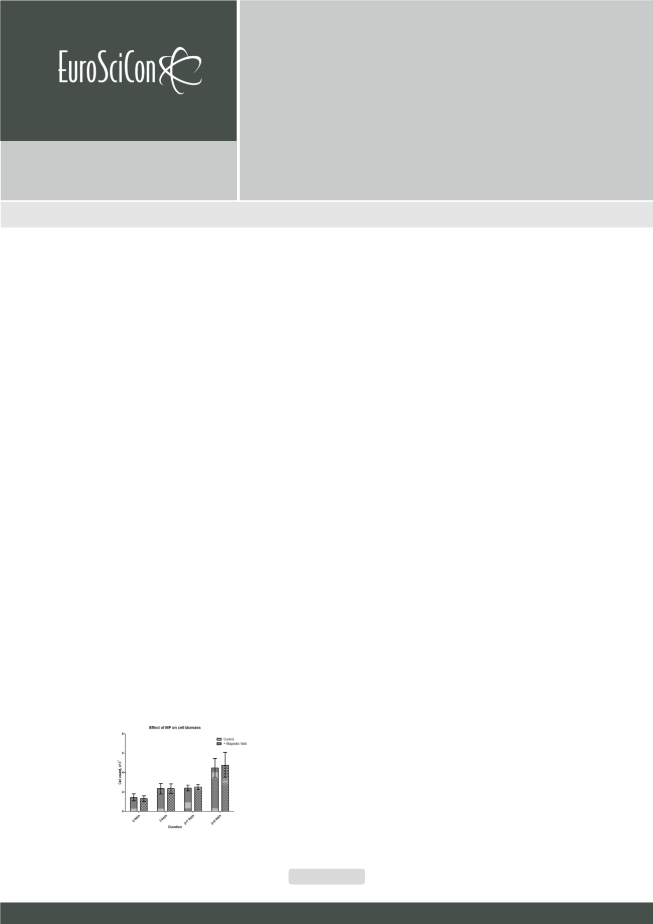
Obtained results showed (Fig.1) that viable cell count after 2-4 days of cultivation varied from 133 to 4816 million.
Magnetic field has no statistically significant influence on cell proliferation. Factors that influence cell proliferation are cultivation
regime and duration. During the fed-batch regime for 4 days, viable cell count increased more than 3 times compared to the batch
regime for 2 days.
Conclusion & Discussion:
The research showed that 0.5T static magnetic field exposure has no statistically significant influence
on CHO cell proliferation. However, proliferation increased due to the change of cultivation regime and duration.
Acknowledgement:
This work has been supported by European Regional Development Fund within the project “Influence of the
magnetic field initiated stirring on biotechnological processes” No. 1.1.1.1/16/A/144
alina.rekena@rtu.lvEvaluation of Magnetic Field Influence on Chinese
Hamster Ovary Cells
A. Rekena
1
, D.Livkisa
2
, D.Loca
1
1
Rudolfs Cimdins Riga Biomaterials Innovations and Development Centre of RTU, Institute of
General Chemical Engineering, Faculty of Materials Science and Applied Chemistry (Riga
Technical University, Pulka 3, Riga, LV-1007, Latvia)
2
Department of Microbiology and Biotechnology, Faculty of Biology (University of Latvia,
Jelgavas 1, Riga, LV-1004, Latvia)
A. Rekena et al., Biochem Mol biol J 2019, Volume:5
DOI: 10.21767/2471-8084-C1-024
Figure 1:
Magnetic field influence on cell proliferation after 2 and 3 days in a batch
regime and 3 and 4 days in a fed-batch regime (2+1 and 2+2 days, respectively).