Localization of Breast Lesion Using Mammographic-Basic Views
Nikki Mishra*, Meena Chakradhar, Ranjit Jha and Rauniyar RK
Department of Radio-Diagnosis and Imaging, B.P. Koirala Institute of Health Sciences (BPKIHS), Dharan, Nepal
- *Corresponding Author:
- Nikki Mishra
Department of Radio-Diagnosis and
Imaging, B.P. Koirala Institute of Health
Sciences (BPKIHS), Dharan, Nepal
Tel: +977-9860735394/ +977-9824381727
E-mail: nikki.mishra438@gmail.com
Received date: May 23, 2018; Accepted date: September 26, 2018; Published date: October 03, 2018
Citation: Mishra N (2018) Localization of Breast Lesion Using Mammographic-Basic Views. Clin Immunol Infect Dis Vol. 2 No. 1: 2.
Abstract
The incidence of breast cancer is more in developed countries (>1000 per million) than in developing countries (<200 per million) but the rate of cancer mortality is higher in developing countries than in developed countries [1]. Cancerous breast lesions accounts for 6% of all cancers in Nepal. This disease is mostly detected in advanced or very late stage due to female illiteracy and lack of awareness in Nepal. Human Female breast is composed of mainly glandular and adipose tissue and has 4 quadrants i.e. upper outer, upper inner, lower outer, lower inner and sub-areolar region. This study was done for two years on females of eastern region of Nepal who visited BPKIHS for mammography examination and at the end of the study it was concluded that Upper outer quadrant of the breast is most susceptible to breast lesion with highest number of pathologies occurring in that area and also mammography was found to be an idealistic imaging modality for both screening and diagnostic purposes of breast
Keywords:
Lesion; Breast quadrant; Mammography
Introduction
Breast cancer is the second most common malignancy among women in Nepal [1]. This disease is mostly detected in advanced or very late stage due to female illiteracy and lack of awareness in Nepal. Factors such as age, menstruation, parity, breast density etc. play a vital role in its occurrence. Localization of lesion in the different breast quadrants may help in the better diagnosis and aid in treatment of the disease which can ultimately lead to the decrease in breast cancer mortality rate in the Eastern region of Nepal. BIRADS, as established by American College of Radiology, stands for Breast Imaging Reporting and Data System which helps in categorizing the lesions and creating a spectrum based upon the nature of lesion whether it is benign or malignant and can also be helpful in predicting lesion that is more frequently occurring in female breasts. Localization of lesion is done using mammography which is both an effective screening and diagnostic tool that helps detect changes in breast up to two years before it appears clinically.
Background
There are many researches done around the globe in order to detect the quadrant of breast that is most susceptible to breast pathologies but none of the significantly related study has been found to be done in Nepal. Therefore, this study was done in BP Koirala Institute of Health Sciences, Nepal, to localize the lesion in different breast quadrants and find the most vulnerable one that is more susceptible to breast lesion using mammographic basic views and also to justify the use of mammography as an affective screening modality for women with increasing age.
Materials and Methods
This was a hospital-based descriptive cross sectional study conducted on 120 female patients in the Department of Radiodiagnosis and imaging, BPKIHS, Dharan, Nepal. Mammography reports of all 120 female patients at age 30 years and above were evaluated during two years study period. A standard questionnaire (including age, weight, height, parity, mammographic indication, menopausal history) was given to the patient to record the clinical history. Mammography was performed on “Planmed Sophie Classic S” scanner machine and findings were classified according to various BIRADS assessment categories (category 0 to category 6). Basic mammographic views (MLO,CC) were taken by standard mammographic techniques ( film focal distance-60 cm, molybdenum target with molybdenum filter with the use of automatic exposure control, compression according to patient tolerance, kVp according to patient habitus). Post-operative breast surgery cases, females with breast implants and male patients were excluded from this study. Each mammogram was evaluated by the concerned radiologist of the department and the findings were noted on structured Performa. Final data was exported to IBM Statistical Package for Social Science Software (SPSS) version 11.5 and analysis was done using simple descriptive statistics. Purposive sampling technique and linear by linear Chisquare test was applied to find the association of lesion with various breast quadrants. P-value less than 0.05 was considered to be statistically significant (confidence level=95%).
Results
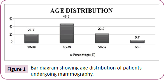
Total 120 cases were evaluated and their age ranged from 30 to 80 years with mean 45.08 and SD +8.861 where maximum patients ranged from the age group 40-49 years. Distribution according to age is summarized in graph below (Figure 1).
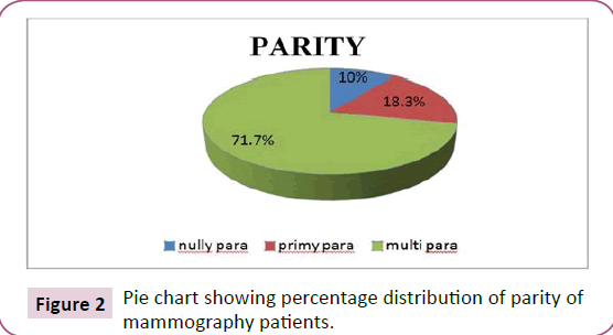
Further classification of patients was done on the basis of parity where majority of the cases were multiparous and the percentage pie-chart is shown below (Figure 2).
Similarly, mammography patients presented with different kinds of clinical indications among which pain was the most common one followed by lump and nipple discharge. Many came for screening purpose and only few came with other clinical features (Table 1). Also the breast tissue of the patient undergoing mammography was evaluated which showed that majority of the patients falling among the age group of 40-49 years had predominantly scattered fibro-glandular tissue (47.5%) and heterogeneously dense (35%) breast tissue whereas other age groups had predominantly fatty and dense breast tissues. The distribution of breast tissue among the patients is shown in Table 2.
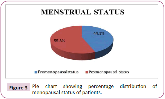
The history of menstruation was taken of all the patients who underwent mammography and was categorized into premenopausal and postmenopausal stages because menstruation plays an important role in hormonal changes within the body that directly affects the breast composition which can later be a reason for cancer development in the breast. Further distribution is shown in the pie-chart below (Figure 3).
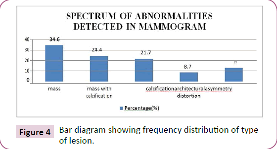
Out of all 120 patients, 71 cases were found normal (BIRADS cat-1) and 49 cases had lesion findings (BIRADS cat-2/3/4/5/6) in different breast quadrants. The results of all the patients who had a lesion (n=49) in their breast were analyzed and further characterization of lesion was done as to whether it was a mass, calcification, architectural distortion or asymmetry. The percentage graph of different abnormalities seen in mammogram is shown below in graph IV (Figure 4).
The distribution of lesion (n=49) was further done in respective right and left breast of the patients and it was found that maximum 25 cases the lesion was present in left breast whereas in 18 cases the lesion was present in right breast of the patients. Only 6 cases had lesion present in both right and left breasts (Table 3).
Finally the above given frequency of lesions were distributed in each quadrants of the right and left breast respectively and the tables are demonstrated below (Tables 4 and 5).
As stated above, we collected total 120 patients of mammography in our department during our study period among which 71 were normal cases whereas 49 were suspected with abnormality (lesion) in their breasts. The lesion characterization was done on the basis of presence of different abnormalities in their mammograms, according to which they were further analyzed into various categories of BI-RADS (Breast Imaging-Reporting and Data System). Among 49, maximum 34 cases were found with benign lesions whereas 15 cases were found with malignant ones and rest of the frequency distribution of patients with lesions and their BI-RADS categories are given in (Table 6).
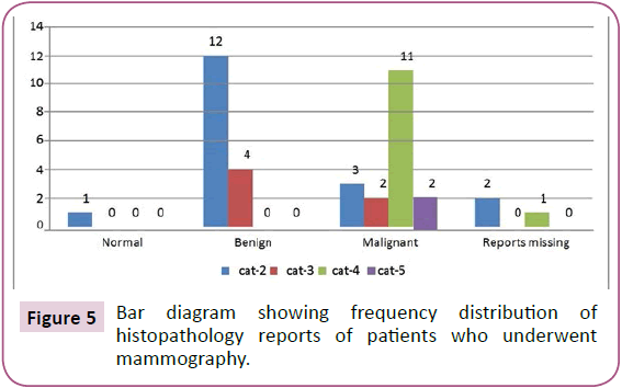
Among total 120 patients, 49 patients were suspected with lesion on their mammograms. Hence, those patients were clinically suggested for histopathologic correlation either through FNAC or biopsy and their final reports were noted. Therefore, out of total 49 cases suspected with lesions in mammography, histopathology reports of 3 patients categorized as cat-2 and cat-4 in mammography were found missing from the laboratory for correlation, 34 cases were found to have histo-pathologically proven lesion be it either benign or malignant, 10 cases that had benign calcification (BIRADS cat-2) in mammogram did not undergo such correlation because mammography is ideal for its confirmation. One patient already had biopsy proven malignancy that was reported as BIRADS cat-6 in mammography (Figure 5).
Discussion
Breast cancer is one of the major causes of death in developing countries like Nepal. Its incidence is progressively increasing due to modern lifestyles and other factors such as age, parity and hormonal changes [2-4]. With the advent of mammography, the diagnosis of breast cancer has significantly improved which facilitates early management and intervention. The vast majority of human malignancies are age-associated cancers. About 80% of all breast cancers arise in women over age 50 [2]. A study done by Singh et al. the commonest age group of patient was 40-50 years which is also similar to the finding noted by Jain et al. where maximum cases were from 41-45 years age group [1,3]. In a study done by Shen et al. the age range was from 24 to 80 years with mean 48.8±9.3 years where the commonest age group was 40 years and above [4]. Similar findings were noted in our study where the age range was 30-80 years with mean 45.0+8.8 years and the commonest age group of patient was 40-49 years.
Delayed child birth i.e. after the age of 30 years is a risk factor for breast cancer. Hormonal factors and reproductive history such as nully parous and first full term pregnancy is associated with increased risk [1]. Parity is an important modulator of breast cancer risk. In a study done by Shen et al. it was found that out of 392 patients, 15 patients were nully parous, 165 cases were primy parous and 212 cases were multiparous which results that the occurrence of breast lesion was least in multiparous patients and it was proved that increased parity was a protective factor against breast cancer [4]. In our study, among 120 patients who underwent mammography, 12 (10%) were nully parous, 22 (18.3%) were primy parous and maximum 86 (71.7%) cases were multiparous with similar association with breast cancer risk and multi parity.
The patients who underwent mammography were symptomatic to certain pathologic conditions like pain, lump, discharge etc. In a study done by singh et al. among 80% of patients presented with painless breast lump, pain and nipple discharge [1]. Similar study was done by Jain et al. where pain was the commonest representation among 120 patients appearing for mammography [3]. Our study showed similar result as Jain et al. where maximum 65 (54.2%) cases showed pain as a common clinical feature of women in this region of Nepal because maximum women are unaware of breast pathologies and are hesitant to reveal. They lack the knowledge of periodic self-examination of breast in their daily life. Most of the women visit any clinician only when they experience any mild or severe pain in their breasts that compel them to seek medical consultancy.
Mammography is more reliable than clinical examination in detecting fatty and fibrous breast tissues. In a study done by Boyd et al. breast density assessed by mammography reflected breast tissue composition where the relationship between breast density and susceptibility to breast cancer was found significantly high [5]. Ahmadinejad et al. did similar kind of research about the association of mammographic density with pathologic findings and he found that out of 131 patients, 9.9% had diffusely fatty breast tissue, 30.5% had scattered fibroglandular tissue, 43.5% had heterogeneously dense tissue and 16% had diffusely dense breast tissue with a conclusion that in patients with high breast density, malignant cases were significantly more in comparison to patients with low breast densities [6]. Similar result was found in our study where out of 120 patients, 12 (10%) cases had diffusely fatty breast tissue, 57 (47.5%) cases had scattered fibroglandular tissue, 42 (35%) cases had heterogeneously dense tissue and 9 (7.5%) cases had diffusely dense breast tissue with occurrence of lesion being more in patients with high breast density.
There are various factors that lead to the onset of breast cancer in women and menopausal status is one of them. In a study done by Benz et al. the age-specific incidence profile was exponential until menopause and increased more slowly thereafter [2]. Similar finding was noted by Seth et al. where the patients detected from benign or malignant lesion were more common in postmenopausal women. Similar result was noted in our study where out of 120 patients, 67 (55.8%) cases were in postmenopausal status whereas only 53 (44.1%) cases were in postmenopausal state with the same increasing risk of breast cancer in postmenopausal women as found by the studies mentioned above.
The cases with lesion findings were further evaluated for the localization of lesion on different quadrants of both right and left breasts. A study done by Darbre, found that the upper outer quadrant is the most common site for breast lesion in women due its high glandularity. The reports of 212,677 patients between the year 1979 and 2000 were evaluated and it revealed that maximum 52.5% of the lesions were in the upper outer quadrant of the breast [7]. Similar result was found in the study done by Rummel et al. where maximum 51.5% of tumors were found to be located in the upper outer quadrant of the breast [8]. Our study found similar results with Darbre P and Rummel S where the association between the occurrence of lesion in upper outer quadrants of each right and left breast was found to be highly significant (p-value=<0.05) where out of 24 lesions in right breast, maximum 15 (62.5%) lesions occurred in upper outer quadrant and similarly, out of 31 lesions in left breast, majority of 20 (64.5%) lesions occurred in upper outer quadrant respectively further proving that upper outer quadrant is the most susceptible area for the occurrence of any type of breast lesions.
Conclusion
We concluded that breast pathologies were more frequent to occur in upper outer quadrant irrespective of any side of breast (right or left) where mass (with or without calcification) seemed to be more frequently occurring lesion in women among 40- 49 years age group. Also, mammography is ideal cost effective choice for patients to detect early breast pathologies.
Limitations
1. False negative results are more common to occur in women with dense breast, hence young women are more prone to have false negative results.
2. Technical faults in machine may produce artifact that might mimic a lesion.
3. Additional views taken to confirm the location of a lesion in a particular breast might have delivered additional dose it.
References
- Singh YP, Sayami P (2009) Management of breast cancer in Nepal. J Nepal Med Assoc 48: 252-257.
- Christopher CB (2008) Impact of aging on biology of breast cancer. Crit Rev Oncol Hematol 66: 65-74.
- Jain SB, Jain I, Shrivastav J, Jain B (2015) A clinic-pathological study of breast lumps in patients presenting in surgery OPD in a referral hospital in Madhya Pradesh, India. Int J Curr Microbiol App Sci 4: 919-923.
- Shen S, Zhong S, Xiao G, Zhou H, Huang W (2017) Parity association with clinicopathological factors in invasive breast cancer: A retrospective analysis. Onco Targets Ther 10: 477-481
- Boyd NF, Martin LJ, Bronskill M, Yaffe MJ, Duric N, et al. (2010) Breast tissue composition and susceptibility to breast cancer. J Natl Cancer Inst 102: 1224-1237.
- Ahmadinejad N, Movahedinia S, Movahedinia S, Shahriari M (2013) Association of mammographic density with pathologic findings. Iran Red Crescent Med J15: e16698
- Darbreh PD (2005) Recorded quadrant incidence of female breast cancer in Great Britain suggests a disproportionate increase in the upper outer quadrant of breast. Anticancer Res 25: 2543-2550.
- Rummel S, Hueman M, Constantino N, Shriver CD, Ellsworth RE (2015) Tumor location within the breast: Does tumour site have prognostic ability? Ecancermedicalscience 9: 552.
Open Access Journals
- Aquaculture & Veterinary Science
- Chemistry & Chemical Sciences
- Clinical Sciences
- Engineering
- General Science
- Genetics & Molecular Biology
- Health Care & Nursing
- Immunology & Microbiology
- Materials Science
- Mathematics & Physics
- Medical Sciences
- Neurology & Psychiatry
- Oncology & Cancer Science
- Pharmaceutical Sciences
