ROLE OF MAGNETIC RESONANCE IMAGING OF BRAIN IN DEVELOPMENTAL DELAY IN CHILDREN
Dibyajyoti Nath, Aabir Hazarika* and Pranjit Thapa
Department of Radio Diagnosis, Silchar Medical College and Hospital, Assam, India
- *Corresponding Author:
- Aabir Hazarika Department of Radio Diagnosis, Silchar Medical College and Hospital, Assam, India, Tel: 8721932881; E-mail:draabir17@gmail.com
Received date: June 23, 2022, Manuscript No. IPCRCR-22-13843; Editor assigned date: June 27, 2022, PreQC No. IPCRCR-22-13843 (PQ); Reviewed date: July 11, 2 022, QC No. IPCRCR-22-13843; Revised date: August 23, 2022, Manuscript No. IPCRCR-22-13843 (R); Published date: August 30, 2022, DOI: 10.36648/IPCRCR.6.5.001
Citation: Nath D, Hazarika A, Thapa P (2022) Role of Magnetic Resonance Imaging of Brain in Developmental Delay in Children. J Clin Radiol Case Rep Vol:6 No:5
Abstract
Introduction: Global developmental delay is diagnosed when there is a significant delay in two or more of the following domains of development: Gross motor, fine motor, speech and language, cognition, and social/ personal development. There are several etiological processes that can hinder the process of developmental of brain in a child such as neurovascular insult, metabolic, toxic and congenital causes among many leading to significant delay in attaining developmental milestones.
Objectives: The purpose of this study is to identify the spectrum of abnormalities of the brain in children presenting with developmental delay on MRI and further categorize the morphological abnormalities.
Materials and methods: Our study included 60 children of both the sexes and age group ranging from 3 months to 12 years with clinical diagnosis of developmental delay referred for brain MRI from the department of paediatrics for a period of one year.
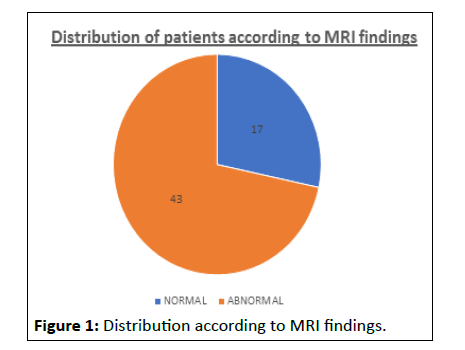
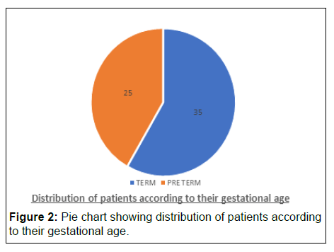
Result: Out of the 60 cases enrolled in the study, 17 (28.3%) patients had normal MRI brain and 43 (71.7%) had abnormal MRI brain findings. The highest number of patients with abnormal MRI brain was found in the age group 3 months-11 months which constituted of 22 patients, followed by age group 1 year–4 years where abnormal brain MRI were found in 15 patients. Out of the total 60 cases, 35 (58.3%) patients had history of term delivery whereas 25 (41.7%) patients had history of preterm delivery. Among the 25 children with history of preterm delivery, 3 (12%) had normal MRI brain finding whereas 22 (88%) had abnormal brain MRI findings. Among the 35 children with history of term delivery, 14 (40%) had normal MRI brain whereas 21 (60%) had abnormal MRI brain findings. Significant association between the gestational age and MRI findings was observed (p<0.05). Among the 43 cases with abnormal MRI brain findings, 33 (53.4%) had findings consistent with neurovascular disease, 9 (15%) cases had congenital anomalies and 2 (3.3%) cases had findings consistent with metabolic disorder. The abnormalities of the ventricles were seen in 25 (41.7%) patients, white matter in 29 patients (48.3%), gray matter in 19 (31.7%) patients, corpus callosum in 20 (33.3%) patients, cerebellum in 6 (10%) patients, basal ganglia in 1 (1.7%) patient and hippocampus in 1 (1.7%) patient.
Conclusion: The MRI brain has been found to be a useful radiological investigation in evaluating a child with developmental delay because it not only allows for a comprehensive assessment of the anatomical detail of the brain, but it also provides an etiological basis for a child with developmental delay, which aids the clinician in providing treatment to the patient and providing counseling to the parents.
Keywords
Magnetic resonance imaging; Developmental delay; Hypoxic ischemic encephalopathy
Introduction
Development is a complex and continuous process of maturity that begins from conception and continues until maturity [1]. Several factors can hinder this process of developmental in a child such as genetic, neurovascular, metabolic, toxic and nutritional among others which can have negative impact and present as significant delay in achieving developmental milestone in a child.
Developmental delay is defined as significant delay (more than two standard deviations below the mean) in one or more developmental domains [2].
Recognizing a fundamental etiology in a child with global developmental delay has applications in treating a potentially curable cause (e.g., metabolic illnesses), evaluating the child's eventual developmental potential [3,4].
Neuroimaging plays an important role in evaluating a child with developmental delay as early neuroimaging curtails other needless investigations, helps in surveillance of known complications, timely initiation of specific intervention and family support [5].
MRI brain helps in assessment of anatomical details of the brain, the presence of any morphological abnormalities and aids the clinician in providing a diagnosis and appropriate treatment in such patients.
Aim and objectives
• To identify the spectrum of abnormalities of the brain in children with developmental delay on MRI.
• To categorize the morphological abnormalities in patients with abnormal brain MRI.
• To identify the proportion of normal MRI in children presenting with developmental delay.
Materials and Methods
Our study included 60 children with clinical diagnosis of developmental delay who were referred for brain MRI to the department of the radiology, Silchar medical college and hospital, Silchar for one year from 01-06-2020 to 31-05-2021. The study population consisted ofb oth the sexes with age ranging from 3 months-12 years.
Inclusion criteria: Patients aged between 3 months to 12 years referred for brain MRI with developmental delay to diagnose the cause are included in the study
Exclusion criteria: Patients less than 3 months. Patients more than 12 years. Patients with general contraindication for MRI. Consent not given for the study.
Magnetic resonance imaging: MRI evaluation was carried out on a Siemens Tim Avanto 1.5T scanner using a head coil under supervision. The procedure and duration of the examination were explained in understandable terms to the parents and informed consent taken. Sedation was performed only for irritable babies under supervision with caution and continuous monitoring of vitals [6]. The older children were either sedated by oral or IV drugs in the presence of the paediatrician and an anesthetist. All children were monitored with ECG and pulse oximetry during the entire scan. Scanning was done in the axial, coronal, and sagittal planes. Sequences used are T1W, T2W, Fluid Attenuation Inversion Recovery (FLAIR), GRE in post gadolinium T1W, Susceptibility Weighted Images (SWI), Diffusion Weighted Imaging (DWI) and Apparent Diffusion Coefficient mapping (ADC Map).
Statistical analysis: Data entered in MS excel sheet and SPSS version 26.0 was used for statistical analysis. Qualitative variables were summarized using frequencies and percentages. Chi square test was used for comparing the qualitative variables. p<0.05 was considered statistically significant [7].
Results
Out of the total 60 cases, brain MRI findings were reported as normal in 17 cases (28.3%) and abnormal findings were seen in 43 cases (71.7%) (Figure 1).
The distribution according to age and MRI findings were studied, the highest numbers of abnormal MRI findings were found in the age group 3 months–11 months which constituted 22 patients, followed by the age group 1 year–4years where abnormal MRI findings were found in 15 patients (Table 1) [8].
| Age | Normal MRI (n=17) | Abnormal MRI (n=43) | ||
|---|---|---|---|---|
| Number | Percentage (%) | Number | Percentage (%) | |
| 3 months-11 months | 6 | 35.3 | 22 | 51.1 |
| 1 year-4 year | 4 | 23.5 | 15 | 34.9 |
| 5 year- 8 year | 4 | 23.5 | 4 | 9.3 |
| 9 year-12 year | 3 | 17.7 | 2 | 4.7 |
Table 1: Distribution according to age and MRI findings.
Out of the total 60 cases, 35 (58.3%) patients had history of were delivery whereas 25 (41.7%) patients had history of preterm delivery (Figure 2).
Out of the 25 children with history of preterm delivery, 3 (12%) had normal MRI finding whereas 22 (88%) had abnormal MRI findings [9]. Among the children with term delivery, 14 (40%) had normal MRI finding whereas 21 (60%) had abnormal MRI findings. There is significant association between the gestational age (preterm delivery) and MRI findings was observed (p<0.05) (Table 2).
| MRI | Pre term | Term | P value | ||
|---|---|---|---|---|---|
| No of patients | Percentage (%) | No. of patients | Percentage (%) | P=0.03 | |
| Normal | 3 | 12 | 14 | 40 | |
| Abnormal | 22 | 88 | 21 | 60 | |
Table 2: Distribution of MRI findings according to the gestational age.
The distribution of affected anatomical structures in patients with abnormal MRI (43 cases) was analyzed. The abnormalities of the ventricles were seen in 25 (41.7%) patients, white matter in 29 patients (48.3%), gray matter in 19 (31.7%) patients, corpus callosum 20 (33.3%) patients, cerebellum in 6 (10%) patients, basal ganglia in 1 (1.7%) patient and hippocampus in 1 (1.7%) patient (Tables 3 and 4).
| Involved brain structures | Abnormal | Normal | ||
|---|---|---|---|---|
| Number | Percentage | Number | Percentage | |
| Ventricles | 25 | 41.70% | 35 | 58.30% |
| White matter | 29 | 48.30% | 31 | 51.70% |
| Gray matter | 19 | 31.70% | 41 | 68.30% |
| Corpus callosum | 20 | 33.30% | 40 | 66.70% |
| Cerebellum | 6 | 10% | 54 | 90% |
| Basal ganglia | 1 | 1.70% | 59 | 96.70% |
| Hippocampus | 1 | 1.70% | 59 | 98.30% |
Table 3: Distribution of involved brain structures.
| Categories | No. of cases | Percentage |
|---|---|---|
| Normal | 17 | 28.30% |
| Neurovascular disease | 32 | 53.40% |
| Congenital and developmental anomalies | 9 | 15% |
| Metabolic disease | 2 | 3.30% |
Table 4: Categorization of MRI findings.
The 60 cases were categorized according to their etiology based on MRI findings. 17 (28.3%) cases had normal MRI findings. Among the 43 cases with abnormal MRI findings, 32 (53.4%) cases had findings consistent with neurovascular disease, 9 (15%) cases had congenital anomalies and 2 (3.3%) cases had findings consistent with metabolic disorder.
The most common abnormality encountered in present study was neurovascular diseases where majority of patient had findings consistent with Hypoxic Ischemic Encephalopathy (HIE) [10]. The total number of patients with HIE in the present study were 31. Majority of these children belonged to 3 month to 11 months of age group. Congenital and developmental anomalies constituted 9 (15%) patients which included:
• Chiari I malformation (1 case)
• Chiari II malformation (1 case)
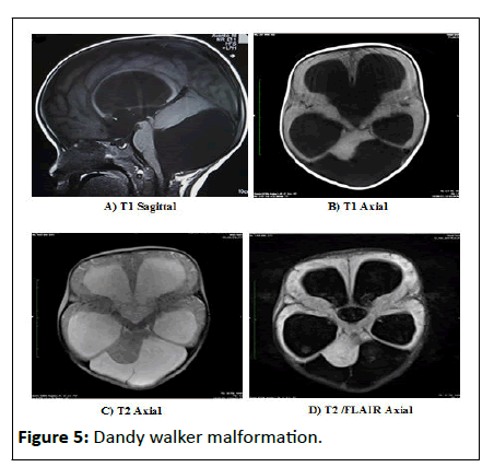
• Dandy walker malformation (4 cases)
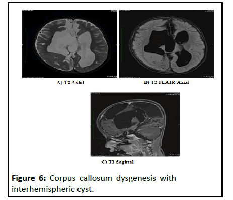
• Corpus callosum agenesis/dysgenesis (2 cases)
• Aqueductal stenosis (1 case)
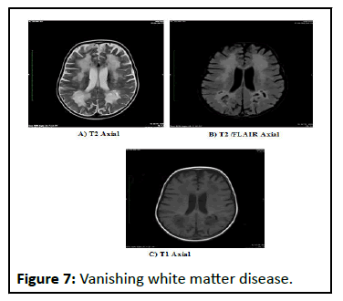
2 patients (3.3%) presented had findings consistent with metabolic encephalopathy which included 1 case with Wilson disease and one case with vanishing white matter disease.
DISCUSSION
In the present study comprising of 60 children, the proportion of children with normal and abnormal MRI brain findings were 28.3% and 71.7% respectively. In the study done by T Arul Dasan and B Deepashree, the proportion of children with an abnormal brain MRI was 72%. Other studies including Abhishek and Kapadia, Anand Mathew, et al., Altaf Ali S, et al. and Momen, et al. had the following percentage of children with abnormal brain MRI 72.5%, 75%, 68% and 58.6% respectively. The findings of our study are comparable to the studies by T Arul Dasan and B Deepashree, Abhishek S and Kapadia and Altaf Ali S, et al.
In the present study, most of the children with abnormal brain MRI findings belonged to age group 3 months to 11 months (51.1%) with the next peak at 1 year-4 year's (34.9%). In the study conducted by Altaf Ali S, et al. 7 of 81 paediatric patients presenting with developmental delay, most of the abnormal MRI findings were in the age group 3-12 months, with the next peak at one to two years. In the study conducted by Momen, et al. 1 of 580 children with developmental disorders, among 56 children aged between 2 and 6 months, 40 (70.59%) had abnormal MRI findings while from 109 children aged 61–164 months, 47 patients (43.12%) had abnormal brain MRI findings. Anand Mathew, et al. in their study of 60 children with developmental disorders found that among the 45 patients with abnormal MRI finding, 20 (71.4%) had abnormal MRI in the under age 2 group. In the 2-5 year's group 19 (86.6%) out of 22 had abnormal MRI and in those above 5 years, 60% of had abnormal MRI findings. T Arul Dasan and B Deepashree in their study of 90 children with developmental delay divided the patients into four age groups: 3 months to 4 years, 5 to 8 years, 9 to 12 years and 13 to 18 years. The frequency of normal brain MRI was higher in the older age groups, whereas the frequency of abnormal brain MRI was found to be the highest in the age group 3 months-4 years (44-48.8%). Hafiz Habibullah, et al. found in their study that out of 170 children diagnosed with GDD, 93 cases had abnormal morphological appearance on MRI and most of the children with abnormal MRI findings were in the age group of 2–5 years (29%), with the next peak at 8–12 years (25.8%). Abhishek S and Kapadia in their study of total 200 children with developmental delay, found maximum number of abnormalities were detected in age group of 5 to 7 years in 26 %of children, followed by 3 to 5 years age in 21.5% of children.
The findings in the present study are comparable with study conducted by Altaf Ali S, et al. and Momen, et al.
In the present study, out of the 25 children with history of preterm delivery, 22 (88%) had abnormal brain MRI findings. Among the children with term delivery, 21 children (60%) had abnormal MRI findings. Significant association between the gestational age (preterm delivery) and MRI findings was observed (p<0.05).
Anand Mathew, et al. in their study of 60 children found that, 16 (26.67%) were preterm deliveries and 44 (73.33%) were term deliveries of the 16 preterm cases 15 (93.7%) had abnormal MRI findings and out of the 44 term cases 30 (68.2%) had abnormal MRI findings. The observed difference was found to have statistical significance between preterm deliveries and abnormal MRI findings (p value=0.04).
The finding of our study is comparable to the study done by Anand Mathew, et al. where statistical significance between preterm deliveries and abnormal MRI finding was found.
In the present study out of the 43 cases with abnormal MRI, white matter (48.3%), ventricles (41.7%) and corpus callosum (33.3%) were the most commonly involved structures. In the study conducted by, Althaf Ali S, et al. the 55 cases with abnormal MRI were evaluated for involvement of various anatomical structures. The ventricles (62%) and corpus callosum (58%) were the most commonly involved structures cases respectively. Widjaja, et al. 11 studied 90 such children and found ventricles (48%) and corpus callosum (44%) were the most commonly involved in the study conducted by Abhishek S and Kapadia, of the 145 cases with abnormal MRI findings the ventricles and white matter in parietal lobe was most common followed by occipital lobe, seen in 29.5% and 17.5 % cases respectively. The finding of our study is similar to Abhishek S and Kapadia where the most common involved structure in patients with abnormal MRI findings was white matter and ventricles.
In the present study 17 (28.3%) cases had normal MRI findings. Among the 43 cases with abnormal MRI findings, 32 (53.4%) cases had findings consistent with neurovascular disease, 9 (15%) cases had congenital anomalies and 2 (3.3%) cases had findings consistent with metabolic disorder. The most common abnormality encountered in present study was neurovascular diseases where majority of patient had findings consistent with Hypoxic Ischemic Encephalopathy (HIE). Majority of these children belonged to 3 months to 11 months of age group. Congenital and developmental anomalies constituted 9 (15%) patients and 2 patients (3.3%) presented had findings consistent with metabolic encephalopathy.
Abhishek and Kapadiya, in their study found that the most common MRI diagnosis of developmental delay was neurovascular insult to the brain found in 59 (29.5%) cases. The second most common MRI diagnosis of developmental delay was structural malformation of brain found in 42 (21%) cases.
Althaf Ali S, et al., in their study found that among the 55 (68%) cases with abnormal MRI findings 25 cases (31%) had findings consistent with traumatic/neurovascular diseases. The most common abnormality encountered in their study was traumatic/neurovascular diseases like Hypoxic Ischemic Encephalopathy (HIE). The congenital and developmental abnormalities of the brain found in 14 (17%) cases. Metabolic and degenerative diseases were found in 8 (10%) cases.
T Arul Dasan and B Deepashree, in their study of 90 patients found that 65 (72%) had abnormal findings on brain MRI. 53 (58.8%) had findings that were categorized as neurovascular diseases and non-specific findings of delayed neurodevelopment. 10 out of 90 (11.1%) patients had structural brain malformations. One (1.1%) male child aged between 5-8 years had tuberous sclerosis. Out of 90 patients 1 had brain MRI findings suggestive of metachromatic leukodystrophy.
Anand Mathew, et al. in their study of 60 children with developmental disorders found that 75% of children had abnormal brain magnetic resonance imaging findings and majority of them had hypoxic ischemic encephalopathy changes (60%), cerebral malformations (6.67%) and nonspecific findings (8.33%). No cases of metabolic causes were identified.
Momen, et al. in their study of 580 children with developmental disorders found that among 340 abnormal MRI cases, 218 (37.6%) had neurovascular diseases.
Armando Freitas da Roch, et al. in their study of 146 children with mental retardation found that the most frequent MRI finding was with structural lesions such as, focal thinning of the corpus callosum at the isthmus level; asymmetry of the lateral ventricles; periventricular leukomalacia; atrophy or dysgenesis of the corpus callosum; congenital malformations.
The findings of our study are similar to the studies done by T Arul Dasan and B Deepashree and Anand Mathew, et al. where the most common abnormality encountered was due to neurovascular disease in 58.8% and 60% respectively comparable to 53.4% of patients in our study. The second most common abnormality in both the studies is due to congenital and structural brain malformation similar to our study.
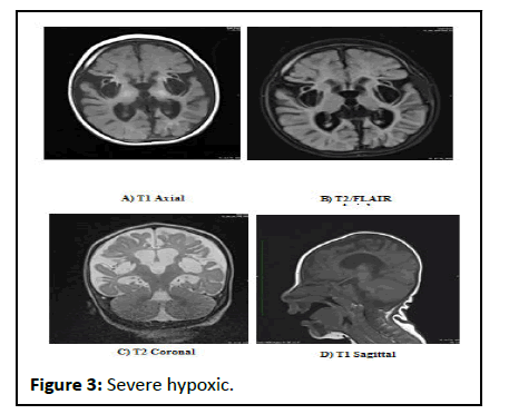
A 5 month old pre term child with a history of birth asphyxia and presenting with history of no neck holding and lack of social smile.
Axial (A) T1W1 (B) T2/FLAIR and (C) T2 Coronal MR images showing cystic encephalomalacic changes in the bilateral parieto temporal region with dilatation of the bilateral lateral ventricles. On sagittal T1WI (D) thinning of the corpus callosum was seen.
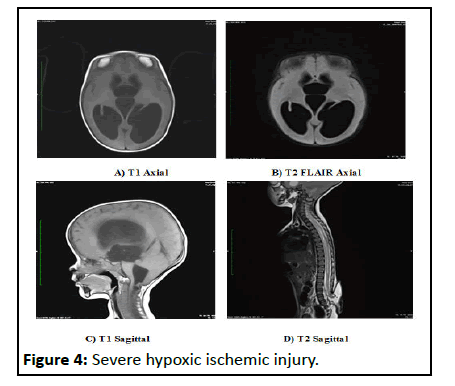
1 year old child presenting w ith developmental delay and seizure.
Axial (A) T1W1 (B) T2/FLAIR MR images showing dilatation of bilateral lateral ventricles and third ventricle. (C) Sagittal T1 MR image showing the cerebellar tonsils and vermis are displaced inferiorly through the foramen magnum. The fourth ventricle is low lying and is elongated [11]. (D) T2 Sagittal image of whole spine showing the presence of lumbo sacral myelomeningocele.
5 month old child presenting with lack of social smile, no neck holding and unable to sit with support.
(A) T1 Sagittal (B) T1 Axial (C) T2 Axial (D)T2/FLAIR Axial MR images showing enlarged posterior fossa, cystic dilatation of the fourth ventricle and hypoplasia of the vermis 7 year old child presenting with mental retardation.
(A) T2 Axial and (B) T2/FLAIR Axial and (C) T1 Sagittal MR Images shows corpus callosal dysgenesis and an interhemispheric cyst which is communicating with right lateral ventricle with dilatation of bilateral lateral ventricles. 11 year old child presenting with developmental delay and mental retardation. A) T2 W1 Axial (B) T2/FLAIR Axial images showing diffuse white matter hyperintensity predominantly involving centrum semi ovale, corona radiata and periventricular region. Axial (A) T2W1 (B) T2/FLAIR (C) T1W1 showing CSF like densities in bilateral periventricular white matter in the parieto-occipital region (Figures 3-7).
Conclusion
Developmental delay present with a wide spectrum of etiologies. MRI is a useful investigation in evaluating such patients especially in children without clinically obvious cause and it helps the clinicians to provide a definitive diagnosis for management of such patients. It is a preferred modality of radiological investigation in such patients as MRI does not use ionizing radiation and depicts anatomy in greater details. The present study could distinguish the normal and abnormal morphological appearances of the brain in such patients and further categorize them into different subgroups. Among the study population the most common cause of abnormal MRI findings was found to be neurovascular where majority of patients had findings consistent with hypoxic ischemic encephalopathy.
Acknowledgements
Authors would like to thank the Department of Radiology, Silchar Medical College and Hospital, Assam, India.
Funding and conflict of interest
None declared
References
- Momen AA, Jelodar G, Dehdashti H (2011) Brain Magnetic Resonance Imaging Findings in Developmentally Delayed Children. Int J Pediatr 2011:1–4
[Crossref] [Googlescholar] [Indexed]
- Battaglia A, Carey JC (2003) Diagnostic evaluation of developmental delay/mental retardation: An overview. Am J Med Genet 117:3–14
[Crossref] [Googlescholar] [Indexed]
- Lemay JF, Herbert AR, Dewey DM, Innes AM (2003) A rational approach to the child with mental retardation for the paediatrician. Paediatr Child Health 8:345-356
[Crossref] [Googlescholar] [Indexed]
- Kjos BO, Umansky R, Barkovich AJ (1990) Brain MR imaging in children with developmental retardation of unknown cause: results in 76 cases. AJNR Am J Neuroradiol 11:1035-1040
[Googlescholar] [Indexed]
- Verbruggen KT, Maurits NM, Meiners LC, Brouwer OF, Van Spronsen FJ, et al. (2009) Quantitative multivoxel proton spectroscopy of the brain in developmental delay. J Magn Reson Imaging 30:716-721
[Crossref] [Googlescholar] [Indexed]
- Abhishek S, Kapadia B (2020) MRI Evaluation of Central Nervous System in Childhood Developmental Delay. Asian J Med Radiol Res 8:1-8
- Ali Althaf S, Syed NP, Murthy GS, Nori M, Abkari A, et al. (2015) “Magnetic resonance imaging (MRI) evaluation of developmental delay in pediatric patients.” J Clin Diagn Res 9:21-24
[Crossref] [Googlescholar] [Indexed]
- Mathew A, Brahmadathan MN, Parvathi R (2022) Brain magnetic resonance imaging in developmentally delayed children a cross sectional study. Cureus 14: 24051
[Crossref] [Googlescholar] [Indexed]
- Habibullah H, Albradie R, Bashir S (2019) Identifying pattern in global developmental delay children: A retrospective study at King Fahad specialist hospital, Dammam (Saudi Arabia). Pediatr Rep 11:8251
[Crossref] [Googlescholar] [Indexed]
- Widjaja E, Nilsson D, Blaser S, Raybaud C (2008) White matter abnormalities in children with idiopathic developmental delay. Acta Radiol 49:589-595
[Crossref] [Googlescholar] [Indexed]
- da Rocha AF, da Costa Leite C, Rocha FT (2006) Mental retardation: a MRI study of 146 Brazilian children. Arq Neuropsiquiatr 64:186-192
Open Access Journals
- Aquaculture & Veterinary Science
- Chemistry & Chemical Sciences
- Clinical Sciences
- Engineering
- General Science
- Genetics & Molecular Biology
- Health Care & Nursing
- Immunology & Microbiology
- Materials Science
- Mathematics & Physics
- Medical Sciences
- Neurology & Psychiatry
- Oncology & Cancer Science
- Pharmaceutical Sciences
