MelatoninâÂÂs Metabolite Ion at m/z 174.1: An Active Biomolecule with the Ability to Reverse the Signs of Skin Aging
Silvia Perrella Segre*, Francesco Di Salvo and Sergio Capurro
Department of Korpocare, Korpo Srl Research, Genoa, Italy
- *Corresponding Author:
- Silvia Perrella Segre
Department of Korpocare,
Korpo Srl Research,
Genoa,
Italy,
Tel: 39010594921;
E-mail:. sergio.capurro@hotmali.it.
Received date: September 25, 2022, Manuscript No. IPJMBB-22-14634; Editor assigned date: September 28, 2022, PreQC No.. IPJMBB-22-14634 (PQ); Reviewed date: October 13, 2022, QC No. IPJMBB-22-14634; Revised date: January 17, 2023, Manuscript No.. IPJMBB-22-14634 (R); Published date January 24, 2023, DOI: 10.36648/ IPJMBB.8.1.002
Citation: Segre SP, Salvo FD, Capurro S (2023) Melatonin’s Metabolite Ion at m/z 174.1: An Active Biomolecule with the Ability to Reverse the. Signs of Skin Aging. J Mol Biol Biotech Vol:8 No:1
Abstract
Background: The treatments to prevent or treat skin. aging are increasing more and more. Consumers are. increasingly choosing skin care products formulated. with active ingredients endowed with great anti-aging. effectiveness. This research has focused on. obtaining melatonin’s metabolite ion at m/z 174.1, a. novel active biomolecule which has the property of. reversing skin aging.
Methods: This manuscript is featuring methods aiming to. generating a large amount of the metabolite ion at m/. z 174.1 possessing high skin anti-aging effective. properties, through ionization and fragmentation of. melatonin.
Results: Melatonin was ionized and fragmented with Far. Infrared Radiation (FIR) and low nergy electrons emitted. from Quantum Dots, structured inside the matrix of a. US-patented silver alloy.
These processes generated various types of metabolites,. including the biologically powerful melatonin metabolite. ion at m/z 174.1.
Conclusion: This extensive research enabled us to discover,. the metabolite ion at m/z 174.1 as an active ingredient. of cosmeceutical bionic serums with the desirable property. of reversing the signs of skin aging.
Keywords
Melatonin; Far Infrared Radiation (FIR);. Fibroblasts; Melatoninergic Antioxidative System (MAS)
Introduction
This research has focused on obtaining melatonin’s. metabolite ion at m/z 174.1, a novel active biomolecule which. has the property of reversing skin aging. Melatonin was ionized. and fragmented with Far Infrared Radiation (FIR) and low-energy. electrons emitted from quantum dots, structured inside the. matrix of a US-patented silver alloy.
These processes generated various types of metabolites,. including the biologically powerful melatonin metabolite ion at. m/z 174.1.
Far infrared radiation and low energy electrons emitted by. quantum dot nanocrystals structured inside the matrix of a. patented silver alloy [1]. AgQDs, can emit Far Infrared Radiation. (FIR) at a wavelength of λ=100 μm. Moreover, because AgQDs. nanocrystals are electrically polarized, they emit a large quantity. of low-energy electrons. QD nanocrystals are in a p-type. form; thus, any electrical energy is conducted, not by the. movement of solution phase ions, but by a hole (electron. vacancy) or electron movement.
Materials and Methods
During FIR, a large portion of the radiant energy carried by. the electromagnetic wave emitted from AgQDs is absorbed. by melatonin, which causes changes in its molecular atomic. and electronic-energy. Upon stimulation by FIR, the energy. level transitions of the p-type QDs are emitted in the. form of electromagnetic radiation. This weak radiation. assists the polarization of melatonin during its ionization.
Melatonin is made of uniquely arranged atoms and molecules. and the molecules move among and between the atoms. When. the molecules are irradiated with a 100 μm wavelength band of. electromagnetic irradiation, the electromagnetic wave energy is. absorbed and the amplitude of melatonin's molecular. vibration is increased. The FIR wavelength of 100 μm was. selected, because it elicits the expected vibrational energy in. melatonin molecules more rapidly and efficiently than other. frequencies and it induces vibrational energy in water molecules. or chains of water molecules, where melatonin is dissolved.
Electron micrographs (magnified 18,000 times) have. demonstrated that c onsiderable nanometer sized. QD nanocrystal clusters are effectively formed with AgQDs.. These minute sized QD nanocrystal clusters result in new. quantum phenomena that yield some extraordinary. advantages during the ionization and fragmentation of. melatonin biomolecules. Moreover, the AgQD surface has. a fractal geometry that improves the FIR energy distribution.
To detect the ionic fragments of melatonin, we used the LCMS/. MS method described by Serban [2]. In short, an Agilent. Zorbax Eclipse XDB C-18 rapid resolution column (50 mm × 4.6. mm ID × 1.8 Μ) was used for the separation of melatonin’s. metabolite ions and detection of the metabolite at m/z 174.1.. The mobile phase A comprised 5 mM ammonium formate in. 80% water and 20% methanol; mobile phase B was 100%. methanol.
Results and Discussion
FIR and low energy electrons emitted by the AgQDs were. activated with thermal energy at 39°C. These electrons impacted. and interacted synergistically with the melatonin molecules. dissolved in a mixture of water and propylene glycol (50/50). As. a result, one electron was lost from the neutral molecule. The. energy of ionization generated by the synergistic action of FIR. and low energy electrons emitted from the AgQDs produced. molecular ions (M+) in an excited state. More precisely, the. melatonin molecule ejected an additional electron, which left a. positively-charged molecular ion of the type:
M+e¯→ M.++2e¯
These ions were formed with a range of internal energies. The. molecular ion obtained during the electron impact decomposed. and formed fragment ions, Ai+ and radicals, Bi..
M+e¯→M.++2e¯→Ai++Bi.
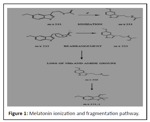
Thus, the fragmentation reaction of the melatonin molecule. took place as shown in Figure 1.
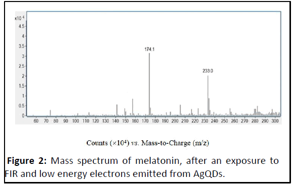
The fragmentation mass spectra of melatonin metabolite ion. at m/z 174.1 is shown in Figure 2.
The specificity of this metabolite could be used as a. fingerprint of the molecular parent, melatonin, which generated. it.
Quantitative determination of melatonin metabolite m/z. 174.1 by LC-MS/MS are described elsewhere.
Many things cause our skin to age. Some things we cannot do. anything about; others we can influence. One thing that. we cannot change is the natural aging process. With time, we all. get visible lines on our face. It is natural for our face to lose. some of its youthful fullness. We notice our skin becoming. thinner and drier. Our genes largely control when these. changes occur. The medical term for this type of aging is. “intrinsic aging.” However, we can influence another type of. aging that affects our skin. Our environment and lifestyle. choices can cause our skin to age prematurely. The medical. term for this type of aging is “extrinsic aging.” By taking some. preventive actions, we can slow the effects of this type of. aging on our skin.
Extrinsic aging occurs because our skin is at the mercy of. many forces as we age: Exposure to the sun (photo-aging), harsh. weather, pollution and bad habits. Many forces cause the. formation of free radicals, which damage cells and lead to,. among other things, premature wrinkles. Other factors that. contribute to wrinkled, spotted skin include the loss of. subcutaneous support (fatty tissue between skin and muscle),. stress, gravity, daily facial movement, obesity and. even sleeping position.
As we discover the fundamental mechanisms that control skin. aging, new anti-aging agents are introduced, such as melatonin.. Melatonin is a hormone produced in the pineal gland that. follows a circadian light dependent rhythm of secretion.. Melatonin receptors are expressed in many skin cell types,. including normal and malignant keratinocytes, melanocytes and. fibroblasts. Melatonin has been experimentally implicated in. skin functions, such as hair cycling and fur pigmentation.. Moreover, it possesses a wide range of endocrine properties and. strong antioxidative activity. The discovery that UV-exposure. induced solar damage to the skin led to the finding that. melatonin could counteract reactive oxygen species generation,. mitochondrial damage and DNA damage. Thus, there was. considerable evidence that melatonin could be an effective antiskin. aging compound.
Early studies detected melatonin metabolites in cell free. systems [3]. More recent studies provided the first evidence of. UV-enhanced photolytic and/or enzymatic melatonin. metabolism in cultured human keratinocytes [4]. Therefore, it was concluded that human keratinocytes and likely the skin and. its appendages, represent an extrapineal site of melatonin. synthesis. Furthermore, these cells could serve as targets for. protective exogenous melatonin treatment. In other words, the. skin possesses fully functioning, local, autonomous. melatonin metabolism [5]. Moreover, all melatonin. metabolites are strongly lipophilic, which enables them to. diffuse readily into skin and cellular compartments.. Because cutaneous local synthesis and metabolism are. inducible by UV-irradiation, it could also be postulated that the. skin possesses a self-regulated protective system that is. switched on by environmental stressors, such as UV. radiation, ionizing radiation and inflammation.
Most investigations of the different aspects of melatonin. metabolites have confirmed that, both biosynthetic. and biodegradation pathways of melatonin are observed in. whole human skin and in the major cutaneous cell. populations. UV-exposure mediates melatonin metabolism and. the generation of melatonin metabolites in human. keratinocytes; these metabolites exert strong antioxidative. properties. Thus, an antioxidative cascade was postulated for. the skin, analogous to previously describe melatonin related. antioxidative cascades in chemical or other tissue homogenate. systems [6]. This cascade was defined as the Melatoninergic. Antioxidative System (MAS) of the skin. The MAS supports. the skin’s function as an important barrier organ by. protecting against UV-induced and oxidative stress-mediated. damaging events, at the levels of the nucleus, subcellular. spaces, proteins and cell morphology.
Therefore, endogenous intracutaneous melatonin production,. together with topically applied exogenous melatonin. metabolites might be expected to represent a potent. antioxidative defense system against UV induced skin. aging. Moreover, it has become increasingly apparent that. melatonin metabolites contribute to the reducing potential of. melatonin. Indeed, it is crucial to understand that the. protective actions of melatonin are not due to melatonin,. per se, but to its metabolites, which can stimulate. antioxidative enzymes [7-9].
Conclusion
In humans, skin aging is accompanied by a decline in. mitochondrial function. The mitochondria serve as the. “powerhouse of the cell” and make 90% of the chemical energy. that cells need to thrive. At the University of Alabama,. Birmingham, researchers have developed a mouse model of. aging, by inducing a mutation in the mouse genome that led to. mitochondrial dysfunction. The mice developed wrinkled skin in. a matter of weeks. When researchers stopped the mitochondrial dysfunction, many of the wrinkles vanished. That. study suggested that mitochondrial decline opened the door. to skin aging.
In a recent study researchers in South Carolina and. Russia showed that melatonin promoted longevity through its. ability to preserve mitochondrial function. Other researchers. found that the natural melatonin hormone worked in a. unique way to combat mitochondrial dysfunction. through i ts various metabolites. Thus, the melatonin. metabolite ion detected at m/z 174.1 is a potent, anti-free. radical, dynamic substance that can preserve mitochondrial. function and thus, reverse the signs of aging.
References
- Tan DX, Manchester LC, Burkhardt S, Sainz RM, Mayo JC, et al. (2001) N1-acetyl-N2-formyl-5-methoxykynuramine, a biogenic amine and melatonin metabolite, functions as a potent antioxidant. FASEB J 15:2294-2296
[Crossref] [Googlescholar] [Indexed]
- Fischer TW, Sweatman TW, Semak I, Sayre RM, Wortsman J, et al. (2006) Constitutive and UV-induced metabolism of melatonin in keratinocytes and cell-free systems. FASEB J 20:1564-1566
[Crossref] [Googlescholar] [Indexed]
- Slominski A, Fischer TW, Zmijewski MA, Wortsman J, Semak I, et al. (2005) On the role of melatonin in skin physiology and pathology. Endocrine 27:137-148
[Crossref] [Googlescholar] [Indexed]
- Tan DX, Manchester LC, Terron MP, Flores LJ, Reiter RJ (2007) One molecule, many derivatives: A never-ending interaction of melatonin with reactive oxygen and nitrogen species? J Pineal Res 42:28-42
[Crossref] [Googlescholar] [Indexed]
- Reiter RJ, Tan DX, Terron MP, Flores LJ, Czarnocki Z (2007) Melatonin and its metabolites: New findings regarding their production and their radical scavenging actions. Acta Biochim Pol 54:1-9
[Googlescholar] [Indexed]
- Singh B, Schoeb TR, Bajpai P, Slominski A, Singh KK (2018) Reversing wrinkled skin and hair loss in mice by restoring mitochondrial function. Cell Death Dis 9:735
[Crossref] [Googlescholar] [Indexed]
- Paradies G, Paradies V, Ruggiero FM, Petrosillo G (2015) Protective role of melatonin in mitochondrial dysfunction and related disorders. Arch Toxicol 89:923-939
[Crossref] [Googlescholar] [Indexed]
- Baburina Y, Odinokova I, Azarashvili T, Akatov V, Lemasters JJ, et al. (2017) 2’,3’-Cyclic nucleotide 3’-phosphodiesterase as a messenger of protection of the mitochondrial function during melatonin treatment in aging. Biochim Biophys Acta Biomembr 1859:94-103
[Crossref] [Googlescholar] [Indexed]
- Kneidinger H, Mitulovic G, Hartmann J, Quint RM, Getoff N (2015) Melatonin free radicals and metabolites resulting by emission and consumption of solvated electrons (eaq-): Reaction mechanisms. In Vivo 29:605-609
[Googlescholar] [Indexed]
Open Access Journals
- Aquaculture & Veterinary Science
- Chemistry & Chemical Sciences
- Clinical Sciences
- Engineering
- General Science
- Genetics & Molecular Biology
- Health Care & Nursing
- Immunology & Microbiology
- Materials Science
- Mathematics & Physics
- Medical Sciences
- Neurology & Psychiatry
- Oncology & Cancer Science
- Pharmaceutical Sciences


