Parietal Peritoneal Hemangioma Mimicking Acute Appendicitis: A Case Report
Subhi Mansour1, Salma Abofoul1, Hany Bahouth1,2, Yuram Kluger1,3 and Safi Khuri1,3*
1Department of General Surgery, Rambam Health Campus, Israel
2Trauma and Acute Care Surgery, Rambam Health Campus, Israel
3Hepatopancreaticobiliary and Surgical Oncology Unit, Rambam Health Campus, Israel
- *Corresponding Author:
- Safi Khuri
Department of General Surgery
Rambam Health Campus, Israel
Tel: +9721700505150
E-mail: s_khuri@rambam.health.gov.il
Received Date: January 25, 2018; Accepted Date: February 05, 2018; Published Date: February 20, 2018
Citation: Mansour S, Abofoul S, Bahouth H, Kluger Y, Khuri S (2018) Parietal Peritoneal Hemangioma Mimicking Acute Appendicitis: A Case Report. Gen Surg Rep. Vol.2 No.1:4
Abstract
Cavernous Hemangiomas, known as Cavernomas, are benign tumors of congenital origin, than can involve any organ site. In the abdominal cavity, the liver is the most affected organ. Abdominal walls Hemangiomas are rarely reported. Herein, we present a case of 20 years old healthy female patient, presented with abdominal pain of acute onset. A diagnosis of acute appendicitis was made by abdominal ultrasonography. During operation, the appendix was without inflammatory signs. A vascular mass originating from the parietal peritoneum at the right lower quadrant was demonstrated. Resection of the mass, along with appendectomy was performed. Pathological exam of the mass was compatible with Cavernous Hemangioma.
Keywords
Vascular; Liver; Pain; Cystic; Cells; Blood; Infection
Introduction
Cavernous Hemangiomas, also known as Cavernous Angiomas or Cavernomas, are defined as vascular malformations or hamartomas of congenital origin. These are benign tumors that arise from unipotent angioblastic cells, and can involve almost all organ sites [1]. They are known to enlarge by ectasia rather than by hyperplasia or hypertrophy [2]. Cavernous Hemangiomas vary in size from a few millimeters to many centimeters. Larger lesion may be pedunculated and on gross examination appear as cystic lesions with a dark color [3]. Microscopically, the tumor is composed of cavernous vascular spaces of varying sizes, lined by a single layer of flat endothelium and filled with blood [4]. Within the abdominal cavity, hemangiomas may occur anywhere, including solid organs, hollow viscera, ligaments, and abdominal wall, with the liver being the most common intra-abdominal organ involved [1]. A review of the current English literature revealed few reported cases of Hemangioma originating in the omentum, gastrosplenic ligament, lesser omentum, and mesoappendix [5]. To the best of our knowledge, abdominal wall Hemangiomas are rarely reported, and parietal peritoneal Hemangiomas have never been reported up to this date. Herein, we present a case of a 20 year old female patient who presented to our emergency department with abdominal pain of acute onset. Abdominal Ultrasonography showed findings compatible with acute appendicitis. During surgery, a mass was demonstrated; Resection of the aforementioned mass along with appendectomy was performed. Histopathologic report was compatible with parietal peritoneal Cavernous Hemangioma and normal appendix.
Case Presentation
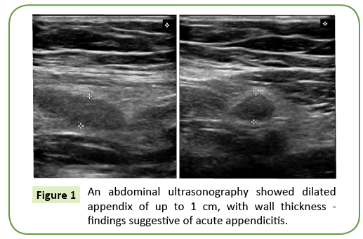
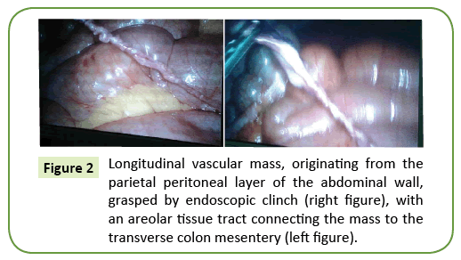
A 20-year-old healthy female patient presented to our emergency department with right lower quadrant abdominal pain of acute onset. The pain lasted for 36 hours, with an acute increase in intensity for the last 8 hours prior to her examination. The pain was accompanied with nausea and anorexia. Upon her admission, her vital signs were within normal limits. The abdominal examination revealed right lower quadrant tenderness with no guarding or rebound. No abdominal mass was palpated. Digital rectal exam was within normal limits. Complete blood count showed normal white blood cell count, liver and kidney function tests were within normal limits. An abdominal ultrasonography (US) showed a dilated appendix of up to a 1 cm, with a thickened wall and increased density of the surrounding fat (Figure 1). These findings were interpreted as acute appendicitis. A gynecologic examination was performed which showed no abnormal findings. The patient was admitted with a diagnosis of acute appendicitis. Due to the above findings, the patient underwent a diagnostic laparoscopy, during which a longitudinal vascular mass, originating from the parietal peritoneal layer of the abdominal wall at the right lower quadrant was demonstrated (Figure 2). An areolar tissue tract connecting the abdominal wall mass to the transverse colon mesentery was also demonstrated. The appendix was without any inflammatory findings. An en bloc resection of the aforementioned abdominal mass and tract tissue along with appendectomy were performed. A close examination of the small and large intestines showed no other origins of inflammation or cause of pain. Her postoperative course was uneventful, and the patient was discharged home on postoperative day 1. The histopathologic examination of the specimen showed parietal peritoneal Cavernous Hemangioma and normal appendix.
Discussion
Hemangiomas are common benign vascular soft tissue tumors affecting mainly adolescents and young adults. The true incidence of these tumors is usually difficult to estimate because they are usually misdiagnosed as venous malformations [6]. Although uncommon, abdominal Cavernous Hemangiomas can occur in the omentum and mesentery because these are derivatives of mesodermal remnants. Hemangiomas are usually associated with nonspecific symptoms. A few reports did mention acute abdomen due to rupture or infection [5]. A case of a 45 yearold woman with a diagnosis of Cavernous Hemangioma arising from the gastro-splenic ligament which presented as acute onset abdominal pain [7]. A similar case reported in which a 62 yearold woman was diagnosed with mesenteric Hemangioma after undergoing two Magnetic Resonance Imaging [8].
Upon reviewing the current English literature we concluded that Cavernous Hemangiomas of the abdominal mesodermal tissues are extremely rare. In all reported cases the diagnosis was made following two or more imaging modalities or during histopathalogic evaluation.
Our patient presented with acute onset of intermittent right lower quadrant pain which mimicked in its clinical presentation and US findings an acute appendicitis. In other cases, these lesions presented as abdominal pain with vomiting, early satiety and weight loss [7], acute abdomen caused by ruptured omental Cavernous Hemangioma [9], ruptured mesoappendix Cavernous Hemangioma [10], infected greater omenal Cavernous Hemangioma [11] or as a palpable mass [12].
The gold standard management for symptomatic abdominal Cavernous Hemangioma is surgical excision. Laparoscopic approach for these lesions is feasible with the advantages of less pain and quicker recovery. After a complete resection, recurrence has never been reported [7].
Conclusion
Cavernous Hemangioma of the mesodermal tissues is a rare tumor that can be challenging to diagnose, and a high index of suspicion is warranted for diagnosis. Once diagnosed, treatment is complete surgical resection of the tumor. So far, no cases of abdominal wall parietal peritoneal Hemangiomas were reported.
References
- Maestroni U, Dinale F, Frattini A, Cortellini P (2005) Ureteral hemangioma: A clinical case report. Acta Biomed 76:115-117.
- Tait N, Richardson AJ, Muguti G, Little JM (1992) Hepatic cavernous haemangioma: A 10 year review. Aust N Z J Surg 62:521-524.
- Baer HU, Dennison AR, Mouton W, Stain SC, Zimmermann A, et al.(1992) Enucleation of giant hemangiomas of the liver. Technical and pathologic aspects of a neglected procedure. Ann Surg 216: 673-676.
- Ishak KG, Rabin L (1975) Benign tumors of the liver. Med Clin North Am 59:995-1013.
- Ojili V, Tirumani SH, Gunabushanam G, Nagar A, Surabhi VR, et al. (2013) Abdominal Hemangiomas: A Pictorial Review of Unusual, Atypical, and Rare Types. Can Assoc Radiol J 64:18-27.
- Priti PS, Siddharth PD, Kaushal C (2014) Anteriorabdominal wall haemangioma with inguinal extension. J Clin Diagn Res 8:ND15-16.
- Kin-Fah C, Ghaith K, Palani SB, David RM (2009) Cavernous hemangioma arising from the gastro-splenic ligament: A case report. World J Gastroenterol 15:3831-3833.
- Takamura M, Murakami T, Kurachi H, Kim T, Enomoto T, et al. (2000) MR imaging of mesenteric hemangioma: a case report.Radiat Med 18:67-69.
- Ritossa C, Ferri M, Destefano I, De Giuli P (1989)Hemoperitoneum caused by cavernous angioma of the omentum. Minerva Chir 44: 907-908.
- Hanatate F, Mizuno Y, Murakami T (1995) Venous hemangioma of the mesoappendix: report of a case and a brief review of the Japanese literature.Surg Today 25:962-964.
- SlizovskiÄ GV, Timashov BA (1995) An infected cavernous hemangioma of the greater omentum in a child. VestnKhirIm. I IGrek 154:69.
- Chateil JF, Saragne-Feuga C, Pérel Y, Brun M, Neuensch wander S, et al. (2000) Capillary haemangioma of the greater omentum in a 5-month-old female infant: a case report. PediatrRadiol 30:837-839.
Open Access Journals
- Aquaculture & Veterinary Science
- Chemistry & Chemical Sciences
- Clinical Sciences
- Engineering
- General Science
- Genetics & Molecular Biology
- Health Care & Nursing
- Immunology & Microbiology
- Materials Science
- Mathematics & Physics
- Medical Sciences
- Neurology & Psychiatry
- Oncology & Cancer Science
- Pharmaceutical Sciences


