ISSN : 2321-2748
American Journal of Phytomedicine and Clinical Therapeutics
Anthocleista Djalonensis Extracts Diminishes Hyperlipidemia through Inhibition of Rho Kinase Expression
Adebayo A Ogunboye1, Olamide O Crown2*, Mary T Olaleye1 and Afolabi C Akinmoladun1
1Department of Biochemistry, School of Sciences, Federal University of Technology, Akure, Nigeria
2Department of Chemistry, Physics and Atmospheric Sciences, Jackson States University, Jackson, Mississippi, USA
- *Corresponding Author:
- Olamide O Crown
Department of Chemistry, Physics and Atmospheric Sciences, Jackson States University, Jackson, Mississippi, USA
E-mail:bayoogunboye@yahoo.co.uk
Received date: 31-Jan-2022, Manuscript No. IPAPCT-22-12302; Editor assigned date: 02-Feb-2022, PreQC No. IPAPCT-22-12302(PQ); Reviewed date: 16-Feb-2022, QC No. IPAPCT-22-12302; Revised date: 23-Feb-2022, Manuscript No. IPAPCT-22-12302(R); Published date: 02-Mar-2022, DOI: 10.36648/2321-2748.10.1.60
Citation: Ogunboye AA, Crown OO, Olaleye MT, Akinmoladun AC (2022). Anthocleista Djalonensis Extracts Diminishes Hyperlipidemia through Inhibition of Rho Kinase Expression. Am J Phytomed Clin Ther Vol.10 No.1:60
Abstract
The effect of Anthocleista Djalonensis Leaf (ADL) extracts on triton-induced hyperlipidemic rats was studied. Fasted experimental rats administered single intraperitoneal (i.p) injections of Triton WR-1339 (200 mg/kg b.w.) were treated with ADL (200, 400, 600 mg/kg b.w.) and Rosuvastatin (10 mg/kg b.w.) except the controls for 14 consecutive days. Serum biochemical indices, expressions of Rho kinase 1 (ROCK 1), β-Hydroxy β-Methylglutaryl-CoA (HMG-CoA) reductase, Geranyl Geranyl Pyrophosphate Synthase (GGPS) and Protein Convertase Subtilisin/Kexin type 9 (PCSK9) in the liver and artery were estimated and the histopathological study of liver and heart were also done. Treatment with the extract significantly ameliorated the great changes produced by triton in serum cholesterol, triglycerides, low density lipoprotein, very low density lipoprotein, atherogenic indices and increased high density lipoprotein and serum albumin levels in a statistically significant manner (p<0.0001). ADL decreased the activities of creatine kinase-MB, gamma glutamyl transpeptidase, lactate dehydrogenase, transaminases and the level of bilirubin in the serum that were elevated by triton while albumin level was increased conversely. Histological examination of the heart and liver showed that the extract markedly protected against hyperlipidemia induced structural distortions. Treatment of animals with ADL and RVT significantly (p<0.0001) down-regulated arterial and hepatic expressions of ROCK 1, HMG-CoA reductase, GGPS and PCSK9 genes. These results indicated that ROCK 1 was expressed upon triton intoxication and leaf extracts from Anthocleista djalonensis was able to mitigate against hyperlipidemia through HMG-CoA reductase activity thereby inhibiting of ROCK1 expression.
Rho kinase 1; β-Hydroxy β-Methylglutaryl-CoA (HMG-CoA) reductase; Geranyl geranyl pyrophosphate synthase; Protein convertase subtilisin/kexin type 9; Hyperlipidemia; Triton; Anthocleista djalonensis; Hyperlipidemia
Introduction
Dyslipidemia contributes majorly to the advent of metabolic syndrome. Metabolic Syndrome (MetS) is a cluster of diseases of metabolism that contributes greatly to increased risk of multiple chronic diseases (such as cardiovascular disease, arthritis, chronic kidney disease, schizophrenia, several types of cancer) and of early death have been reported. As it is clear, MetS puts every individual at risk for atherosclerotic cardiovascular disease. Dyslipidemia is known to play a significant role in the development of atherosclerosis [1].
The most common type of dyslipidemia is hyperlipidemia. Hyperlipidemia is characterised by an abnormal increase in lipid levels i.e. elevated serum level of triglycerides, cholesterol and LDL in the biological system. About a decade ago, the development of lipid lowering drugs or formulation from natural sources have gained importance in such a way that the use of many lipid lowering conventional drugs is being restricted due to several considerable side effects. The triton-induced hyperlipidemia model is popularly utilized in the screening of natural or chemical hypolipidemic drugs. The non-ionic surfactant interferes with the uptake of plasma lipids and has been widely used to block clearance of triglyceride-rich lipoproteins to induce acute hyperlipidemia in several animals.
Atherosclerosis is characterized by progressive inflammation, lipid accumulation and fibrosis in arterial walls. Rho/ROCK pathway is substantially involved in inflammatory and arteriosclerotic arterial lesions in animals and humans. Multiple studies in experimental models involving fasudil and other ROCK inhibitors have demonstrated that ROCK is a critical contributor to the inflammatory atherosclerotic process [2].
Several inhibitors of Rho kinase have been synthesized but only one (Fasudil) has made its way to the bedside. The role of Rho kinase in patients with dyslipidemia and essential hypertension was investigated with treatment with statins and antihypertensive agents, which improved Rho kinase activity [3]. Statins are a class of drugs known for their anti-lipid (HMG-CoA reductase inhibitor) and anti-inflammatory properties. It was reported to have a pleiotropic role in ROCK inhibition. Scientific reports have revealed that cholesterol synthesis activates geranyl geranylation which in turn activates ROCK. Statins inhibit HMG-CoA reductase (rate limiting step in cholesterol biosynthesis) which inhibits indirectly ROCK which therefore prevents activation of apoptosis, angiogenesis, and others. In addition, it preferably inhibits small GTP-binding proteins, such as Rho, isoprenylation rather than lipid lowering. With isoprenylation, small GTP-binding proteins such as Rho can affect endothelium, leukocyte, and fibroblast in different ways. Under pathological conditions, such as hyperlipidemia and diabetes mellitus, Rho isoprenylation and its effector Rho kinase (ROCK) negatively affect the vascular system. As a result, there will be a promotion in inflammatory cells adhesion and infiltration, impairing endothelial function, increasing reactive oxidative generation, destabilizing eNOS mRNA and decreasing NO production [4].
Anthocleista djalonensis A. Chev (Loganiaceae) is a medium-sized tree of the West tropical Africa, 30 feet–45 feet high with blunt spines on the unbranch, pale grey trunk and wide spreading crown. Ethnomedicinally, the seeds, barks, leaves and roots are used as antipyretic, laxative, remedy for various stomach disorders, antihelmintic, antimycobacterial, antibacterial and wound healing [5]. It is used as herbal remedy for epilepsy. The methanolic extract of A. djalonensis leaves showed a very potent DPPH and O2- anion radical scavenging activities. Also, the extract displayed significantly higher OH radical and non-enzymatic lipid peroxidation inhibitory potentials than that of standard antioxidants. It also inhibited the accumulation of nitrite in vitro[6]. However, little is known about the antihyperlipidemic potential and its pharmacological mechanisms. Hence, this study seeks to investigate the effect of ADL on Rho kinase expression in triton induced hyperlipidemia.
Materials and Methods
Materials
Chemicals and reagents: All chemicals were of analytical grade. Marker enzymes and lipid profile kits were obtained from Randox laboratories (Antrim, Northern Ireland, UK). Triton WR-1339 was obtained from Scharlau (Scharlab S.L, Spain). All other chemicals used in the experiment were of the highest grade commercially available.
Plant material: Collection and identification: Leaves of Anthocleista djalonensis were collected from Yasere village in Okitipupa (6°25’ and 6°25’ N latitude and 4°35’ and 4°50’ E longitude), Nigeria. The plant was authenticated by Mr. Omoomoh Bernard of department of forestry and wood technology, federal university of technology, Akure, Nigeria herbarium with a voucher number FUTA-0150. The air-dried leaves were subjected to a coarse powder using a dry grinder. The powdered leaves were soaked in 80% methanol for 72 hours and filtered using Whatman filter paper no.1 to obtain the Aqueous Methanolic Extract (ADL). The filtered extracts were concentrated in a rotary evaporator and further concentrated to dryness using a freeze dryer. The residue was kept at -20°C for further studies.
Animals: The male Wistar rats weighing 190 g ± 20 g were obtained from animal care facility, university of Ibadan, Ibadan. The animals were left to acclimatize for two weeks and kept under a standard environmental condition of 12 hours light and dark, before the commencement of the assay. The standard protocol the national institutes of health guide for the care and use of laboratory animals (NIH Publications No. 8023, revised 1978) was carefully followed. The experimental animals used was approved by the animal and ethics committee of the federal university of technology FUTA 0150.
Hyperlipidemia was induced according to [7]. Briefly, male Wistar rats weighing 190 g ± 20 g were divided into seven groups of seven animals each. All animals were fasted for 18 h and hyperlipidemia prior to induction of hyperlipidemia and grouped as follows: Group I (control) was given the vehicle (i.p), groups II-groups VI was given: Triton (200 mg/kg b.w.), while group VII administered the vehicle. After the intraperitoneal injection of triton X-1339/vehicle, The animals were fasted for 24 h, groups II-VI were treated with Anthocleista djalonensis (200, 400, 600 mg/kg b.w.), and Rosuvastatin (10 mg/kg) b.w.), respectively while groups 1 and VII were normal saline and Anthocleista djalonensis alone(400 mg/kg) b.w.) for 14 consecutive days. The experiment was terminated by sacrificing the rats and blood samples were collected and spinned at 3000 g for 15 min at 25°C to obtain serum. The clear supernatants collected were stored at -20°C for further estimation of biochemical parameters.
Methods
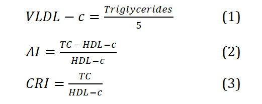
Biochemical assays and atherogenic indices: Serum Aspartate Aminotransferase (AST), Alanine Aminotransferase (ALT), albumin, bilirubin (total and direct) Lactate Dehydrogenase (LDH) and Creatine Kinase (CK) and Gamma Glutamyl Transferase (GGT) activity were estimated using the manufacturer’s procedure (Randox laboratories, UK). Lipid profiles (Total Cholesterol (TC), Triglycerides (TG), Low Density Lipoprotein-Cholesterol (LDL-c), and High-Density Lipoprotein–Cholesterol (HDL-c) were estimated using assay kits supplied by Randox laboratories UK following the manufacturer’s instructions. LDL-c/HDL-c ratio was determined and Very Low-Density Lipoproteins (VLDL-c) [8], Atherogenic Index (AI) and Coronary Risk Indexes (CRI) [9] were calculated as follows:
Histopathological analysis: Histopathological assessment was carried out according to [10]. Briefly, the excised heart and liver were fixed in 10% solution of neutral buffered formalin (pH 7.4). After processing, the tissue was embedded in paraffin and the sections of 5 μm were taken using microtome and stained with hematoxylin-eosin. The slides were examined under a light microscope and the magnified images of the tissue structures were captured.
Gene expression study
RNA extraction: The RNA was extracted as previously described by Lawal et al. Total RNA was extracted from the liver and artery with TRIzol reagent (Thermo Fisher Scientific). The isolated RNA was reconstituted with nuclease free water and quantified spectrophotometrically (A and E Lab UK) at optical density 260 nm/280 nm. The purity of the isolated RNA was calculated from the ratio of absorbances at 260 nm and 280 nm. Calculated absorbance values of RNA between 1.8 and 2.2 were considered as pure RNA samples (Wilfinger et al., 1997) and were used for downstream expressions.
cDNA synthesis: Isolated RNA (1 µg) was reversed transcribed as oligo-dT priming using ProtoScript II First Strand cDNA synthesis kit (Biolabs, New England) in a 3-step reaction condition: 65°C, 42°C for and 80°C for 5 min, 1 hr, and 5 min respectively.
Gene expression analysis by RT-Qpcr: Real-time PCR amplification reactions were performed using OneTaq® 2X Master Mix (NEB) the manufacturer's protocols. Primers for real time PCR reactions were purchased from Inqababiotec (Hatfield, SA) primer sequences are listed in Table 1 (Tables 1 and 2).
| Gene | Sequence |
|---|---|
| PCSK9 | Forward: GCTTCAGCGGCTTGTTCCT |
| Reverse: TGCTCCTCCACTCTCCACATAA | |
| HMGCoA reductase | Forward: TGCTGCTTTGGCTGTATGTC |
| Reverse: TGAGCGTGAACAAGAACCAG | |
| ROCK 1 | Forward: TGCAAGCGCAATTGGTAGAAG |
| Reverse: AATGCAGGCAGAACCAACTG | |
| GGPPS | Forward: TGGTGTGTAGAACTGCTCCAGGCTTTCTTC |
| Reverse: ACACTGGGGTCTCCAAAGAGATCAAGGTAG |
Table 1: The relative amount of cDNA was quantified by Comparative Cycle Threshold (ΔΔCT) method and gene expression was normalised with GAPDH gene as housekeeping gene.
| Treatments/ | Control | Triton | TRI+ADL (200 mg/kg) | TRI+ADL (400 mg/kg) | TRI+ADL | TRI+RVT | ADL |
|---|---|---|---|---|---|---|---|
| parameters | (600 mg/kg) | (10mg/kg) | (400mg/kg) | ||||
| CK- MB(U/l)x102 | 5.99 ± 0.65 | 1.43 ± 0.83# | 1.22 ± 0. 96* | 8.92 ± 0. 33* | 5.80 ± 0.85* | 9.25 ± 0.34* | 6.45 ± 0.53* |
| GGT (U/l) | 2.64 ± 1.29 | 14.39 ± 2.20# | 7.78 ± 1.29* | 4.47 ± 1.56* | 2.43 ± 0.82* | 2.32 ± 1.33* | 2.18 ± 0.90* |
| ALT (U/l) | 0.58 ± 0.00 | 2.87 ± 0.20# | 2.22 ± 0.10* | 1.64 ± 0.10* | 1.21 ± 0.08* | 1.57 ± 0.08* | 1.52 ± 0.10* |
| AST (U/l) | 1.28 ± 0.00 | 6.38 ± 0.20# | 4.92 ± 0.20* | 3.17 ± 0.20* | 2.59 ± 0.19* | 3.17 ± 0.20* | 2.98 ± 0.20* |
| ALB (U/l) | 3.09 ± 0.04 | 2.38 ± 0.09# | 2.17 ± 0.20* | 3.03 ± 0.04* | 3.19 ± 0.07* | 2.97 ± 0.04* | 3.25 ± 0.03* |
| Total Bil. (U/l) | 0.21 ± 0.03 | 1.03 ± 0.06# | 0.63 ± 0.05* | 0.37 ± 0.03* | 0.21 ± 0.05* | 0.32 ± 0.03* | 0.24 ± 0.02* |
| Direct Bil.(U/l) | 0.68 ± 0.17 | 1.97 ± 0.06# | 2.67 ± 0.10* | 1.26 ± 0.07* | 0.89 ± 0.13* | 1.53 ± 0.13* | 1.20 ± 0.07* |
| LDH (U/l) x102 | 9.65 ± 0.64 | 13.76 ± 0.75# | 11.12 ± 1.32* | 8.01 ± 0.32* | 4.83 ± 0.80* | 5.93 ± 0. 42* | 8.68 ± 0.24* |
| TC(mg/dl) | 64.17 ± 1.29 | 96.17 ± 1.30# | 79.92 ± 0.53* | 55.67 ± 0.87* | 46.74 ± 1.04* | 75.11 ± 2.44 * | 48.14 ± 1.38* |
| TRIG(mg/dl) | 81.01 ± 0.86 | 224.01 ± 6.46# | 108.65 ± 0.55* | 76.09 ± 1.34* | 43.18 ± 4.52* | 67.97 ± 1.23* | 41.89 ± 2.59* |
| HDL (mg/dl) | 23.62 ± 0.95 | 9.36 ± 0.95# | 13.19 ± 0.50* | 24.44 ± 0.95* | 32.25 ± 0.48* | 31.09 ± 0.55* | 26.75 ± 0.55* |
| LDL (mg/dl) | 24.06 ± 1.89 | 41.93 ± 1.34# | 45.00 ± 1.18* | 18.21 ± 0.94* | 5.92 ± 0.50* | 30.43 ± 1.39* | 13.13 ± 1.65* |
| VLDL (mg/dl) | 16.20 ± 1.35 | 44.79 ± 3.67# | 21.73 ± 1.06* | 15.19 ± 0.67* | 8.64 ± 0.90* | 13.63 ± 0.94* | 8.38 ± 0.52* |
Table 2: Effects of Anthocleista djalonensis leaf extract post-treatment on some serum biochemical parameters on Triton X-1339 induced hyperlipidemic rats. Results are expressed as mean ± SD (n=7); #p<0.0001 vs. control; *p<0.0001 vs. triton; TRI: Triton; ADL: Anthocleista Djalonensis Leaf; TRI: Triton; RVT: Rosuvastatin; ALT: Alanine Aminotransferase; AST: Aspartate Aminotransferase; LDH: Lactate Dehydrogenase; ALB: Albumin, BIL: Bilirubin; GGT = Gamma Glutamyl Transferase; CK-MB = Creatine Kinase-MB; results are expressed as mean ± SD (n=7); ####p<0.0001 vs. control; ****p<0.0001 vs. triton; TC: Total Cholesterol; TRIG: Triglycerides; HDL: High Density Lipoprotein; LDL: Low Density Lipoprotein; VLDL: Very Low Density Lipoprotein.
The size and purity of the amplicon generated from the qPCR assay was further analyzed using 0.8% agarose (prepared in 1 × tris base, glacial acetic acid, EDTA, TAE, buffer). The samples and quick-load purple 100 bp DNA ladder (Biolabs, New England) were loaded on the ethidium bromide stained gel and run at 100 mA/gel. The gel was visualized under ultraviolet light and photographed. The intensities of the bands from agarose gel electrophoresis were quantified densitometrically using Image J software. Representative snapshot of reverse transcription polymerase chain reaction-agarose gel electrophoresis data was plotted as a bar graph.
Polymerase Chain Reaction (PCR): The assay was performed using an optimization template (cDNA) 5 μl, nucleas-free water 3.5 μl, forward primers 2 μl and reverse primers 2 μl and master mix 2 μl. Reverse transcription-PCR reaction was performed in a 15.0 μl final volume. Amplification conditions were: 94°C pre-denaturation for 5 min, 94°C for 30 s, annealing 55°C for 30 s and Extension 72°C for 30 s and then 5 min at 72°C by 30 cycles [11].
Gel electrophoresis: Assessment of polymerase chain reaction products (amplicons) were electrophoresed in 0.2% of agarose gel using 0.5 × TBE buffer (2.6 g of Tris base, 5 g of Tris boric acid and 2 ml of 0.5 M EDTA and adjusted to pH 8.3 with the sodium hydroxide pellet) with 3 μl EZ-vision (VWR life science). The expression product was visualized as bands by blue-light-transilluminator. The intensities of the bands from agarose gel electrophoresis were was quantified densitometrically using Image J software. Representative snapshot of reverse transcription polymerase chain reaction-agarose gel electrophoresis data was plotted as a bar graph [11].
Statistical Analysis
Statistical analysis was performed using one way Analysis of Variance (ANOVA) followed by Tukey post-hoc multiple comparison tests. GraphPad Prism 6.01 (GraphPad Software Inc., CA, USA) statistical software used for data analysis and data was expressed as mean ± standard deviation. In all the tests, p<0.05 was taken as criterion for statistical significance.
Results
Serum lipid profile parameters in Wistar rats
The statistically significant increases in plasma concentration of TC, TG, LDL-c and VLDL-c (P<0.0001), coupled with the significant decrease in HDL-c in triton-intoxicated rats, when compared with the normal control (Table 3), Anthocleista djalonensis significantly (P<0.0001), attenuated triton-induced alterations observed in lipid profile. However, 200 mg/kg b.w of ADL had no significant effect on the alterations induced in LDL-c on the overall. The activity of ADL compared well with Rosuvastatin against triton.
| Group | CRI | AI |
|---|---|---|
| Control | 2.74 ± 0.46 | 1.76 ± 0.45 |
| TRI (200mg/kg) | 10.50 ± 1.22#### | 10.37 ± 2.81#### |
| TRI+ADL(200mg/kg) | 6.36 ± 1.83**** | 5.36 ± 1.83**** |
| TRI+ADL(400mg/kg) | 2.43 ± 0.36**** | 1.43 ± 0.36**** |
| TRI+ADL(600mg/kg) | 1.46 ± 0.10**** | 0.46 ± 0.10**** |
| TRI+RVT(10mg/kg) | 2.44 ± 0.31**** | 1.44 ± 0.30**** |
| ADL(400mg/kg) | 1.81 ± 0.23**** | 0.73 ± 0.26**** |
Table 3: Effects of Anthocleista djalonensis leaf extract post-treatment on atherogenic indices of triton X-1339 induced hyperlipidemic rats. Results are expressed as mean ± SD (n=7); ####p<0.0001 vs. control; ****p<0.0001 vs. triton; TRI: Triton; ADL: Anthocleista Djalonensis; RVT: Rosuvastatin; CRI: Coronary Risk Index; AI: Atherogenic Index
Serum biochemical parameters of Wistar rats
The mean activities of serum CK-MB, GGT, AST, ALT, bilirubin (total and direct), and LDH were found to be significantly (p< 0.0001) higher in triton-induced hyperlipidemic rats than those in control. Anthocleista djalonensis and Rosuvastatin caused a marked decrease in activity at the evaluated dosages. Serum albumin activities was found to be (p<0.01; p<0.0001) significantly lower in triton-induced rats when compared with the control, ADL and Rosuvastatin groups. The results showed that ADL compared well with Rosuvastatin in reversing the cardiac and hepatic damage especially at the highest dosage (600mg/kg) caused by triton (Table 2).
Atherogenic indices
The injection of triton caused significant (P < 0.0001) elevation on Coronary Risk Index (CRI) and Atherogenic Index (AI), and ADL treatment significantly ameliorated the alteration caused by triton compared with control (Table 3).
Histopathological analysis
Plates 1 and 2 show sections of heart and liver of experimental animals after administration of triton and post treatment Anthocleista djalonensis and Rosuvastatin. 1a control (vehicle) group: Normal morphology and architecture with no lesion; 1b triton-induced, hyperlipidemic group: Severe necrosis of the myocyte and fatty degeneration; 1c (TRI+ADL200 mg/kg): Necrosis and mild inflammation of myocyte; 1d (TRI+ADL400 mg/kg): Very mild perivascular inflammation; 1e (TRI+ADL600 mg/kg): Very mild infiltration and inflammation of myocardium; 1f (TRI+RVT10 mg/kg): Mild infiltration and inflammation of myocardium. 2a control (vehicle) group: Normal hepatocytes, sinusoids, central veins and portal triads and no significant lesion; 2b triton-induced, hyperlipidemic group: Fat vacuolation, necrosis and inflammation of the periportal cells; 2c (TRI+200 mg/kg): Mild periportal inflammation; 2d (TRI+400 mg/kg): Mild periportal inflammation; 2e (TRI+600 mg/kg): Moderate infiltration of inflammatory cells; 2f (TRI+RVT10 mg/kg): moderate infiltration of inflammatory cells. The results demonstrated that the histopathological changes induced in the cardiac and hepatic cells of Triton WR-1339 treated rats were markedly reduced in the rats post treated with Anthocleista djalonensis and Rosuvastatin (Figures 1 and 2).

Figure 1: Light microscopic architecture in the heart section stained with Hematoxylin and Eosin (H&E): ×100 TRI = Triton; ADL: Anthocleista Djalonensis Leaf; RVT: Rosuvastatin.

Figure 2: Light microscopic architecture in the hepatic section stained with Hematoxylin and Eosin (H&E): × 100; TRI: Triton; ADL: Anthocleista Djalonensis; RVT: Rosuvastatin.
Effects of methanolic leaf extract of Anthocleista djalonensis on gene expression in artery and liver of triton-induced Wistar rats
The results showed that there was a significant (p<0.0001) increase the activity of Rho kinase 1 in the triton-induced group when compare with the control. Anthocleista djalonensis and Rosuvastatin lower significantly (p<0.0001) the Rho kinase 1 activity in a dose-dependent fashion in the artery when compared with the induced group. Furthermore ADL at higher dose (600 mg/kg) was not able to compete with the standard drug Rosuvastatin. The results showed the activity of Rho kinase 1 in the liver. There was a significant (p<0.0001) increase in the activity Rho kinase 1 in the triton-induced group when compare with the control. Administration of Anthocleista djalonensis and Rosuvastatin lower significantly (P<0.0001) the Rho kinase 1 activity in the different treatment groups when compared with the induced group (Figures 3-6).

Figure 3: Effect of compounds identified from Anthocleista djalonensis on HMGCoA reductase expression in Triton-induced hyperlipidemia in rats using RT-PCR analysis.

Figure 4: Effect of compounds identified from Anthocleista djalonensis on Rho kinase 1 expression in triton-induced hyperlipidemia in rats using RT-PCR analysis.

Figure 5: Effect of compounds identified from Anthocleista djalonensis on PCSK9 mRNA expression in triton-induced hyperlipidemia in rats using RT-PCR analysis.

Figure 6: Effect of compounds identified from Anthocleista djalonensis on GGPP mRNA expression in triton-induced hyperlipidemia in rats using RT-PCR analysis.
Snapshot representation of RT-PCR and chain reaction-agarose gel electrophoresis was carried on GGPP gene followed by densitometric analysis. Data are expressed as mean ± SEM of 7rats (n=7); ####P<0.0001 vs. control; *P<0.05 vs. triton; ***p<0.001 vs. triton; **p*p<0.0001 vs. triton; ADL: Anthocleista Djalonensis aqueous methanolic extract; TRI: Triton; RVT: Rosuvastatin.
Discussion
Metabolic disorders as a result of hyperlipidemia is the most common cause of some degenerative diseases such as cancer, cardiovascular diseases etc. [12]. Though, increase in the blood lipids is not a recent or short-lived phenomenon; yet, it has been rampant in recent times due to technological innovation and increase in urbanization which has caused a lot of drift in our lifestyles. The alarming cases of hyperlipidemia have attracted serious attention of researchers and health workers across the globe. Sadly, the therapeutic options that are available in the management of hyperlipidemia have not measured up to the incidences currently on ground. Hence, prevention of hyperlipidemia will be of the essence in the management of metabolic disorders and its associated complications.
Triton is one of the drugs employed in mimicking the alteration of lipid levels in animal models. In the present study, alterations in the levels of lipids coupled with liver and cardiac toxicity by triton was largely evident and in line with previous studies. Dyslipidemia is common with liver because it is the major metabolic organ in the human system, and it is stressed during pathology.
Plant derived natural products are being explored in search of new therapeutics. Several researchers have reported the role of plants in the management of hyperlipidemia which is in agreement with our findings [13]. In this study, treatment with Anthocleista Djalonensis Leaves Extract (ADL) lessened dyslipidemic condition occasioned by triton toxicity in experimental rats.
Sometimes, elevated serum HDL level may occur following the inhibition of 3-hydroxy-3-methylglutarylcoenzyme A (HMG-CoA) reductase. Although, the mechanism(s) of anti-hyperlipidemia activities of the plant is still unknown. However, its stimulating effect on lipoprotein lipase activity have been suggested to possess some similarities with that of Rosuvastatin. This theory may explain the significant percentage reduction in triglyceride, LDL and VLDL concentration. Also, the observed low level of HDL in the hyperlipidemic rats is consistent with earlier studies [14]. In the current investigation, ADL treatment decreased the levels of total cholesterol and triglycerides, and increased the levels of HDL, thereby suggesting its lipid lowering potential. The hypolipidemic effect of the plant may be due to flavonoids and/or saponins which we found to be part of the main constituents of the study plant. Also, the lipid lowering role of the extract could be attributed to the fact that it contains constituents that are able to inhibit enzymes such as hydroxyl-methyl-glutaryl-CoA (HMG-coA) reductase which participates in the de novo synthesis of cholesterol [15]. Cholesterol and triacylglycerols are of great significance in cardiovascular integrity since high levels of triglyceride and LDL-c are major coronary risk factors [16]. The atherogenic implication of this is that cholesterol in the arterial wall originates principally from LDL-c, which can be isolated from human arterial tissue in amounts directly related to its concentration in serum.
Elevation of CK-MB, GGT, ALT, AST and LDH activities in the serum of triton treated rats indicate that triton may have induced liver injury compared to the control rats. Injury to liver tissues due to hyperlipidemia alters their transport functional capacity and membrane permeability or rather integrity, leading to leakage of the enzymes from the hepatocytes cells [17]. Therefore, the marked release of AST, ALT, and GGT into the circulation indicates severe damage to the hepatic tissue membranes. However, post-treatment of rats with ADL caused a significant (p<0.0001) reduction in the elevated activities of the evaluated marker enzymes. The effect of the extract was dose dependent and it compared favorably with the activity of the standard drug (Rosuvastatin). Detection of cardiac injury could be afforded by measuring serum levels of known cardiac marker enzymes such as CK-MB, AST, LDH and serum lipid profile [18]. Serum creatine kinase activity is a more sensitive indicator of early stage of myocardial ischemia whereas LDH levels give a rough estimate of the extent of injury to myocardial tissues [19]. GGT has been reported to be a very strong risk factor for all forms of heart diseases and a possible indicator for early development of atherosclerosis. Since the levels of these cellular enzymes present in blood are directly related to the intactness of the plasma membrane of the cardiac cells; then, the dose-dependent inhibition of the serum marker enzymes by ADL could be alluded to its role in the maintenance of cardiac membrane integrity and subsequent restriction of enzyme leakages [20]. who performed intravascular sonography and correlated plaque lipid profile fractions and serum LDL with CK-MB levels proved that CK-MB levels were significantly correlating with severity of the Coronary Artery Disease (CAD) and serum LDL levels. Also, [21] reported that CK-MB elevation correlates with coronary artery plaque burden and calcification which are considered to be the principal causes for myocardial ischemic lesions. ALT, AST and LDH are notable markers of hepatocyte injury; however, LDH is less specific. LDH activity has also been reported to increase in ischemic liver injury, myocardial or pulmonary infarction, kidney, heart, liver, lungs and skeletal muscle damage [22]. The increase in the serum activities of these enzymes were directly proportional to the degree of cellular damage caused by hypercholesterolemia. Meanwhile, the activities of these enzymes were elevated in the serum confirming the liver damage. Hence, the reduction of ALT, AST and LDH activities in the extracts treated group suggested that the extracts may protect against ischemic liver or hepatocyte injury. GGT is an enzyme that is an independent risk factor in cardiovascular mortality. This indicates that the higher the elevation of GGT, the greater the risk of death. Besides, increased GGT activity or prognostic index have been linked to chronic forms of coronary heart disease, congestive heart failure and ischemic or hemorrhagic stroke. However, the reduction in serum GGT activity following post-treatment with ADL further suggested the cardio protective potentials of the extract. Though, the exact mechanism of action(s) of ADL is unknown but the hypolipidemic effect may have been facilitated by its constituent phytochemicals such as flavonoids [23]. The effect of triton-induced hyperlipidemia on total bilirubin, direct bilirubin and albumin revealed a significant (p<0.0001) increase in the serum concentration of total and direct bilirubin coupled with significant (p<0.0001) reduction in albumin level in the hyperlipidemic rats when compared with the control rats. However, serum from hyperlipidemic rats that were treated with ADL significantly (p<0.0001) exhibited declined bilirubin (total and direct) and elevated albumin concentrations; a similar result was observed for the rats that were treated with the standard drug. The reduction in serum albumin concentration and elevated bilirubin may be due to hypercholesterolemia-induced liver damage occasioned by abnormal elevation in the activities of hepatic proteins. It may also be as a result of excessive hemolytic destruction of erythrocytes, obstruction of the biliary tract (obstructive jaundice), and drug-induced reactions. The observed reduction in the albumin may be associated with liver damage and subsequent treatment with ADL and Rosuvastatin might have prevented liver damage by improving the albumin/bilirubin balance in the serum.
Furthermore, atherogenic risk predictor indices (CRI and AI) of triton intoxicated rats significantly increased when compared to that of the rats in the control group. These indices are critical in the clinical setting for assessing the risk of cardiovascular diseases beyond the routinely done lipid profile. The positive relationship between LDL-c, and CRI and AI in this study is largely conspicuous, and in tandem with one of our previous studies [24]. On the other hand, ADL and RVT did not only ameliorate the effect of triton but diminished it. It is interesting to note that, ADL at 400 mg/kg compared favorably with RVT while 600 mg/kg had a better therapeutic potential on all the parameters earlier discussed. These findings suggested that ADL could be a possible therapeutic potential in the management of abnormal lipid profile and related cardiovascular diseases.
In the present study, the observed marked attenuation of triton-induced oxidative stress by ADL was further corroborated by the results from our histopathological assessment on cardiac and hepatic tissues from the experimental rats (plates 1 and 2). Cardiac sections from normal control rats showed normal morphology and architecture with no lesion (plate 1a) whereas the triton-induced group had severe necrosis of the myocyte and fatty degeneration (plate 1b). Post treatment with various doses of Anthocleista djalonensis (plates 1c-1f) and Rosuvastatin showed considerable recovery patterns marked with mild necrosis and inflammation of myocyte as well as extremely mild infiltration and inflammation of the perivascular vessels. On the other hand, sections of liver tissues from control rats showed no observable lesion; characterised with normal hepatocytes, sinusoids, central veins (plate 2a),while hepatic tissues of the triton-intoxicated rats showed significant histopathological alterations as adjudged by obvious fat vacuolation, necrosis and inflammation of the periportal cells (plate 2b). However, post treatment with various dose of ADL and RVT showed reduced histological alterations when compared with the induced group (plates 2c-2f). In general, the structural derangements manifesting as severe necrosis, edema, loss of striation and destruction of the endothelial cells of the heart and liver tissues were significantly corrected in rats treated with ADL and RVT.
It is well accepted that enzymes are the major regulators of the lipid metabolism; wherein HMG-CoA reductase, Rho kinase and PCSK9 are some of the most clinically important enzymes involved in the cholesterol biosynthetic pathway [25]. Changes in the activities of these enzymes are closely related to changes in the overall rate of cholesterol synthesis; thus, suggesting that the inhibition of these enzymes would be an effective means to lower serum cholesterol. Rho-associated kinases (ROCKs) belong to the AGC family of serine/threonine kinases. The strong interest in the Rho-ROCK pathway for drug targeting is based on the observation that the abnormal activation of this pathway plays a crucial role in numerous and diverse human diseases such as tumor invasion, angiogenesis, and metastasis, cardiovascular disorders such as coronary vasospasm, cerebral cavernous malformation, hypertension, atherosclerosis, pulmonary hypertension, cardiac hypertrophy, and stroke, insulin resistance, metabolic diseases, and diabetic nephropathy and neurodegenerative disorders [26]. In this study, the arterial and hepatic Rho kinase 1 gene expressions in triton-induced hyperlipidemic rats increased significantly (P<0.0001) compared with control. Treatment of hyperlipidemic rats with ADL and Rosuvastatin significantly (P<0.0001) reduced the Rho kinase 1 activity. In addition to inhibiting cholesterol synthesis, statins also block the synthesis of isoprenoid intermediates such as Farnesyl Pyrophosphate (FPP) and Geranyl Geranyl Pyrophosphate (GGPP) [27]. Both FPP and GGPP serve as important lipid attachments for the post translational modification of a variety of proteins, including heterotrimeric G proteins and small GTP-binding proteins belonging to the family of Ras, Rho, Rap, and RabGTPases [28]. Isoprenylation is critical for intracellular trafficking and function of small GTP-binding proteins. In general, modification with FPP is necessary for proper localization of Ras family proteins, whereas GGPP is required for Rho, Rab, and Rap family proteins. However, some Rho GTPases require both farnesylation and geranyl geranylation for proper function and intracellular localization. By inhibiting mevalonate synthesis, statins inhibit the synthesis of isoprenoid intermediates thereby preventing isoprenylation of small GTPases, leading to the inhibition of these signaling molecules [29]. The ability of ADL to reduce the activity of Rho kinase may be due to its abilities to down regulate HMG-CoA reductase activity which is the rate limiting step in cholesterol biosynthesis vis-à-vis the intermediate isoprenylation that produces Ras, Rho, Rap and RabGTPases. Also Rho kinases contribute to atherogenesis through inflammation, oxidative stress and plaque formation causing atherosclerosis. This is because Rho kinase 1 and 2 play a vital roles in the organization of smooth and non-smooth muscles, cytoskeleton and adhesion plaques as well as transcription factors regulating cellular motility, morphology, polarity, and gene expression. Therefore, down regulating Rho kinase 1 expression by ADL and RVT, is one of the ways to prevent atherosclerosis vis-à-vis inflammation and lipid accumulation. It is proposed that ADL may exert its antihyperlipidemic effect through the aforementioned mechanisms. This is the first time activation of ROCK by triton will be reported. This activation can be explained in the figure below (Figure 7).

Figure 7: The First time activation of ROCK by triton.
Activation of ROCK1 is directly proportional to the expression of geranyl-geranyl pyrophosphate synthase. Metabolically the action of ROCK1 lies at the point that eventually paves way for GGPP activation. Note worthily, lipid peroxidation is an event in acute hyperlipidemic condition [30] and the model used for induction of hyperlipidemia was able to alter other parameters of antioxidant defense showing evidence that oxidative stress is an early event in hyperlipidemia. Herein, triton increases the production of free radicals by enhancing mitochondrial respiration and down regulating the antioxidant system [17]. The increased levels of triglycerides and cholesterol in blood are associated with mitochondrial dysfunction since it induces an overproduction of superoxide radicals possibly due exacerbated xanthine oxidase activity in the endothelium which causes oxidative alterations on their lipids, proteins, and DNA. In addition, circulating LDL oxidized by free radicals are major culprits in cardiovascular complications [31].
Furthermore, findings from this study revealed that the expression of PCSK9 was increased by triton intoxication. PCSK9 regulates the levels of LDL-receptor, i.e. increase in PCSK9 is directly proportional to decrease in LDL receptors [32]. It could be inferred from this that increased expression of PCSK9 could be associated the elevated LDL level as earlier reported in this study. Free radicals oxidize circulating LDL which made it to attach to the arterial wall thereby causing atherosclerosis [31] and eventually cardiovascular events. Rho kinase is a threonine-serine kinase reported to be activated in several diseases in which hyperlipidemia is not an exception. Activation of these pathways could be as a result of oxidative stress or through synthesis of other important isoprenoid intermediates of the cholesterol biosynthetic pathway, such as Farnesyl Pyrophosphate (FPP) and Geranyl-Geranyl Pyrophosphate (GGPP) that are downstream from l-mevalonic acid.
Conclusion
In conclusion, it could be said that the methanolic leaf extract of Anthocleista djalonensis exhibited a significant hypolipidemic activity and an ability to attenuate the accelerated development of atherosclerosis in hypercholesterolemic models.
References
- Zhang C, Adamos C, Jin oh M, Baruah J, Ayee MAA, et al. (2017) OxLDL-induced endothelial proliferation via Rho/ROCK/Akt/p27kip1 signaling: prevention by cholesterol loading. Am J Physiol Cell Physiol: 340-351. [Crossref], [Google scholar], [Indexed]
- Matsumoto A, Manthey HD, Marsh SA, Fassett RG, de Haan JB, et al. (2012) Effects of exercise training and RhoA/ROCK inhibition on plaque in ApoE(−/−) mice. Int J Cardiol 21. [Cross ref], [Google scholar]
- Kajikawa M, Noma K, Maruhashi T, Mikami S, Iwamoto Y, et al. (2014) Rho-associated kinase activity is a predictor of cardiovascular outcomes. Hypertension 63: 856. [Cross ref], [Google scholar]
- Liu AG, Ford NA, Hu FB. Zelman KM, Mozaffarian D, et al. (2017) A healthy approach to dietary fats: Understanding the science and taking action to reduce consumer confusion.Nutr J 16: 53. [Cross ref], [Google scholar]
- Esimone CO, Nworu CS, Onuigbo EB, Omeje JU, Nsirim KL, et al. (2009) Anti-mycobacterial activity of root and leaf extracts of Anthocleista djalonensis (Loganiaceae) and Diospyrosmes piliformis (Ebenaceae). Int J Green Pharm 3: 201-205. [Cross ref], [Google scholar]
- Awah FM, Tufon E, Uzoegwu PN, (2010) Free radical scavenging activity and phenolic contents of Anthocleista djalonensis (Loganiaceae) leaf extract. Int J Biol Chem Sci 4: 2314–2323. [Cross ref], [Google scholar]
- Crown OO, Komolafe TR, Akinmoladun AC, Olaleye MT, Akindahunsi AA, et al. (2018) Parinari curatellifolia seed flavonoids protect against triton‐induced dyslipidemia and atherogenicity in rats.Traditional & Kampo Medicine 5: 11-18. [Cross ref], [Google scholar]
- Friedwald WT, Levy RI, Fredrickson DS (1972) Estimation of the concentration of low-density lipoprotein cholesterol in plasma, without the use of the preparative ultracentrifuge. Clin Chem 18: 499-502. [Cross ref], [Google scholar]
- Del Bas JM, Fernández-Larrea J, Blay M, Ardèvol A, Salvadó MJ, et al. (2005) Grape seed procyanidins improve atherosclerotic risk index and induce liver CYP7A1 and SHP expression in healthy rats. FASEB J 19: 479-481. [Cross ref], [Google scholar], [Indexed]
- Sikarwar VS, Zhao M, Clough P, Yao J, Zhong X, et al. (2016) An overview of advances in biomass gasification. Energy Environ Sci 29: 2939–77. [Cross ref], [Google scholar]
- Elekofehinti OO, Ariyo EO, Akinjiyan MO, Olayeriju OS, Lawal AO, et al. (2018) Potential use of bitter melon (Momordica charantia) derived compounds as antidiabetics: in silico and studies. Pathiophysiology 25: 327–333. [Cross ref], [Google scholar], [Indexed]
- Global Burden of Disease Study Collaborators (2015) Global, regional, and national incidence, prevalence, and years lived with disability for 301 acute and chronic diseases and injuries in 188 countries, 1990-2013: A systematic analysis for the Global Burden of Disease Study 2013. GBD 386: 743-800. [Cross ref], [Google scholar]
- Srivastava R, Srivastava P (2018) Lipid lowering activity of some medicinal plants: A Review of Literature. Biomed J Sci Tech Res 9. [Cross ref],
- Mohammed GJ, AL-Jassani MJ, Hameed IH, (2016) Antibacterial, antifungal activity and chemical analysis of punica grantanum (pomegranate peel) using GCMS and FTIR spectroscopy. Int J Pharmacogn Phytochem 8: 480-494.
[Cross ref], [Google Scholar]
- Islam A, Asadujjaman M, Hossain MS, Morshed MTI, Azizur MR, et al. (2011) Antihyperlipidemic and hepatoprotective effects of different fractions of brassica oleracae in alloxan induced diabetic rats. Int J Pharm Sci Res 2: 1662-1668. [Cross ref],[Google scholar]
- Singh AK, Singh R (2016) Triglyceride and cardiovascular risk: A critical appraisal.Indian J Endocrinol Metab20: 418–428. [Cross ref], [Google Scholar]
- Abdou HM, Yousef MI, Newairy AA (2018) Triton WR-1339-induced hyperlipidemia, DNA fragmentation, neurotransmitters inhibition, oxidative damage, histopathological and morphometric changes: The protective role of soybean oil. JOBAZ 79: 51. [Crossref], [Google Scholar]
- Habte ML, Melka DS, Degef M, Menon MKC, Yifter H, et al. (2020) Comparison of Lipid Profile, Liver Enzymes, Creatine Kinase and Lactate Dehydrogenase Among Type II Diabetes Mellitus Patients on Statin Therapy.Diabetes MetabSyndrObes 13: 763-773. [Crossref], [Google Scholar], [Indexed]
- Aydin S, Ugur K, Aydin S, Sahin İ, Yardim M (2019) Biomarkers in acute myocardial infarction: Current perspectives.Vasc Health Risk Manag. 15: 1-10. [Cross ref], [Google Scholar], [Indexed]
- Uetani T, Amano T, Ando H, Yokoi K, Arai K, et al. (2008) The correlation between lipid volumes in the target lesion, measured by integrated backscatter intravascular ultrasound, and post-procedural myocardial infarction in patients with elective stent implantation. Eur Heart J 29: 1714–20. [Cross ref], [Google Scholar]
- Mehran R, Dangas G, Mintz GS, Lansky AJ, Pichard AD, et al. (2000) Atherosclerotic plaque burden and CK-MB enzyme elevation after coronary interventions: Intravascular ultrasound study of 2256 patients. Circulation 101: 604–10. [Crossref], [Google Scholar], [Indexed]
- Nader S, Ghasem S, Fakher R, Abolfazl S, Soheila N (2011) Modulating effect of soy protein on serum cardiac enzymes in cholesterol-fed rats. Int J Med Med Sci 3: 390-395.
[Cross ref], [Google Scholar]
- Olorunnisola OS, Bradley G, Afolayan AJ (2011a) Antioxidant properties and cytotoxicity evaluation of methanolic extract of dried and fresh rhizomes of Tulbaghia violacea Afr J Pharm. Pharmacol 5: 2490-2497. [Crossref],[Google Scholar]
- Manuwa TR, Akinmoladun AC, Crown OO, Komolafe K, Olaleye MT (2017) Toxicological assessment and ameliorative effects of parinari curatellifolia alkaloids on triton-induced hyperlipidemia and atherogenicity in rats. Proc Natl Acad Sci India B Biol Sci 87: 611–623.
[Crossref], [Google Scholar], [Indexed]
- Silbernagel G, Steiner LK, Hollstein T, Fauler G, Scharnagl H, et al. (2019) The interrelations between PCSK9 metabolism and cholesterol synthesis and absorption.Journal of lipid research, 60: 161-167. [Crossref], [Google scholar], [Index]
- Richardson K, Schoen M, French B, Umscheidm CA, Mitchell MD, (2013) Statins and cognitive function: A systematic review. Annals of Internal Medicine. 159: 688–97. [Crossref], [Google scholar], [Indexed]
- Parihar SP, Guler R, Brombacher F (2019) Statins: A viable candidate for host-directed therapy against infectious diseases.Nat Rev Immunol, 19: 104–117. [Cross ref], [Google scholar]
- Pinner AL, Mueller TM, Alganem, K, McCullumsmith R, Woodruff JHM (2020) Protein expression of prenyl transferase subunits in postmortem schizophrenia dorsolateral prefrontal cortex.Transl Psychiatry, 10: 3. [Crossref], [Google scholar]
- Gbelcová, H, Rimpelová S, Knejzlík Z, Šáchová J, Kolář M, et al. (2017) Isoprenoids responsible for protein prenylation modulate the biological effects of statins on pancreatic cancer cells.Lipids Health Dis. 16: 250. [Cross ref], [Google scholar]
- Ahn HY, Cho HD, Cho YS (2020) Anti-oxidant and anti-hyperlipidemic effects of cordycepin-rich Cordyceps militaris in a Sprague–Dawley rat model of alcohol-induced hyperlipidemia and oxidative stress.Bioresour Bioprocess 7: 33. [Cross ref], [Google scholar]
- Marventano S, Kolacz P, Castellano S, Galvano F, Buscemi S, et al. (2015) "A review of recent evidence in human studies of n-3 and n-6 PUFA intake on cardiovascular disease, cancer, and depressive disorders: Does the ratio really matter?" International Journal of Food Sciences and Nutrition 66: 611-622. [Crossref], [Google scholar], [Indexed]
- Tang Y, Li SL, Hu JH, Sun KJ, Liu LL, et al. (2016) Research progress on alternative non-classical mechanisms of PCSK9 in atherosclerosis in patients with and without diabetes.Cardiovasc Diabetol 19: 33.

Open Access Journals
- Aquaculture & Veterinary Science
- Chemistry & Chemical Sciences
- Clinical Sciences
- Engineering
- General Science
- Genetics & Molecular Biology
- Health Care & Nursing
- Immunology & Microbiology
- Materials Science
- Mathematics & Physics
- Medical Sciences
- Neurology & Psychiatry
- Oncology & Cancer Science
- Pharmaceutical Sciences