A Riddle of Esophagography: Where Do the Descending Aorta and the Abdominal Aorta Run in Cases of the Right Aortic Arch? Report of two cases
1Department of General Medicine, Shonan Keiiku Hospital, Kanagawa, Japan
2Department of Preventive Medicine, Sanno Medical Center, Tokyo, Japan
3Center for Preventive Medicine, Chemotherapy Institute Kaken Hospital, Chiba, Japan
- *Corresponding Author:
- Sugiura Y
Department of General Medicine
Shonan Keiiku Hospital
4360 Endo Fujisawa 252-0816, Kanagawa, Japan
Tel: +81-466-48-0050
E-mail: sugiurayoshi@outlook.com
Received date: September 14, 2018; Accepted date: October 28, 2018; Published date: November 02, 2018
Citation: Sugiura Y, Shigematsu H, Miyata T, Tani N, Morishita T, et al. (2018) A Riddle of Esophagography: Where Do the Descending Aorta and the Abdominal Aorta Run in Cases of the Right Aortic Arch? Report of two cases. J Clin Radiol Case Rep. Vol.2 No.1:06.
Copyright: © 2018 Sugiura Y, et al. This is an open-access article distributed under the terms of the Creative Commons Attribution License, which permits unrestricted use, distribution, and reproduction in any medium, provided the original author and source are credited.
Abstract
Background: The right aortic arch is a rare anomaly of the aorta and is seen at 0.03~0.04% of the population. Case Presentation: We evaluated upper gastrointestinal series for 4,000 examinees between 2012 and 2016 and encountered two cases of the right aortic arch on esophagography.
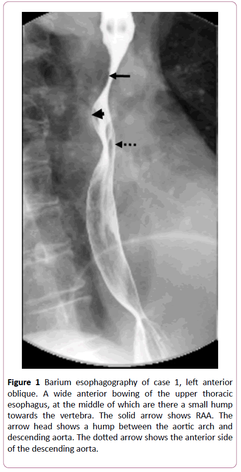
Case 1: Barium esophagography of a seventies-year-old Japanese female showed a wide anterior bowing of the upper thoracic esophagus, at the middle of which was there a small hump towards the vertebra.
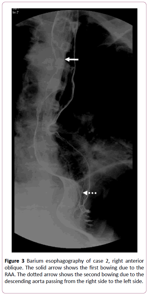
Case 2: Barium esophagography of a forties-year-old Japanese male showed at the first anterior bowing of the upper thoracic esophagus and then an anterior bowing of the lower thoracic esophagus. In the present cases, there adjacently are the upper thoracic vertebrae and upper thoracic esophagus, however, the right aortic arch runs across in between. Where do the descending aorta and the abdominal aorta run? We analyzed barium esophagography. In case 1, aright aortic arch runs at the upper bowing and the descending aorta soon returns from the right side of vertebra to the left side of the vertebra. In case 2, right aortic arch runs in case 1 as well, however, the descending aorta returns to the left above the diaphragm. It was suggested that the aorta in cases of right aortic arch be the shape of a right-side-left question mark.
Conclusion: The descending aorta should be considered to run from the right side to the left side of the thoracic vertebrae.
Keywords
Abdominal aorta; Descending aorta; Right aortic arch
Abbreviations
RAA: Right aortic arch; UGIS: Upper gastrointestinal series; REAA: Retroesophageal aortic arch; US: Ultrasonography.
Introduction
In Japan a health check mainly for company persons has been long time prevailed. It is called “human dock” as if a big ship every several years after on trial has to sleep in a dry dock for her body check. At Sanno Medical Center and Chemotherapy Institute Kaken Hospital, the department of preventive medicine, we accepted 20,000 medical examinees a year; one third of them have upper gastrointestinal series (UGIS) for a gastrointestinal check. The first author evaluated UGIS for 4,000 examinees excluding repeaters between 2012 and 2016 and found 2 cases of the right aortic arch (RAA) on barium esophagography. RAA is seen at 0.03~0.04% of the population [1]. There adjacently are the upper thoracic vertebrae and upper thoracic esophagus, however, the RAA runs across in between.
Case presentation
Case 1
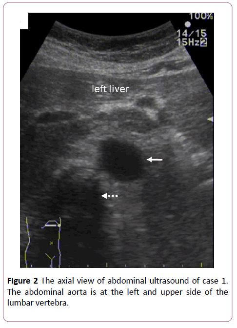
Barium esophagography of a seventies-year-old Japanese female showed a wide anterior bowing of the upper thoracic esophagus, with no symptoms of swallow disturbance, at the middle of which was there a small hump towards the vertebra. An anterior bowing above the hump is considerable due to the RAA. An anterior bowing below the hump should be considered to be the descending aorta running from the right side to the left side of the thoracic vertebrae. The lower thoracic esophagus was never lifted. This case happened to have an abdominal ultrasonography (Figure 1). The axial view showed the abdominal aorta at the left and upper side of the lumbar vertebra (Figure 2).
Figure 1: Barium esophagography of case 1, left anterior oblique. A wide anterior bowing of the upper thoracic esophagus, at the middle of which are there a small hump towards the vertebra. The solid arrow shows RAA. The arrow head shows a hump between the aortic arch and descending aorta. The dotted arrow shows the anterior side of the descending aorta.
Case 2
Barium esophagography of a forties-year-old Japanese male showed at the first anterior bowing of the upper thoracic esophagus and then again an anterior bowing of the lower thoracic esophagus (Figure 3). He had no symptom of swallow disturbance. The first bowing seemed due to the RAA. The second bowing might be due to the descending aorta passing from the right side to the left side. Neither computed tomography nor US was taken in this case as he was a healthy examinee.
Both chest scout films were typical for RAA, hardly showing situs inversus.
Discussion
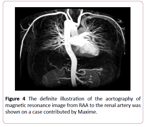
Tons of papers of RAA have been published so far; however, they were much focused on topography in which branches of the arch are discussed. It is clinically useful for cardiovascular, pulmonary and gastroenterological surgeons. For each instance, it was useful to determine the design in endovascular treatment for Kommerell’s diverticulum including intimal tear of the dissection [2]. In a case of the RAA with left lung cancer, a surgeon must pay attention to the path way of the left recurrent laryngeal nerve for mediastinal lymphadenectomy [3]. Successful resection of esophageal cancer with RAA was performed by video-assisted thoracoscopic surgery [4]. The trachea and esophagus were encircled by RAA, the aortic diverticulum and the pulmonary artery. On the paper, barium esophagography showed the indentation of the upper thoracic esophagus at the RAA resembling our esophagography, but not describing how the lower thoracic esophagus was. More exactly our cases belong to the retroesophageal aortic arch (REAA). Philip et al. described it further dividing the right or left REAA. The right REAA has right ascending and left descending aorta with retroesophageal segment, whereas the left REAA has left ascending and right descending aorta. Four of eight cases were symptomatic. Our two cases adjusted the REAA [6]. Finally, the macroscopic view of RAA was illustrated on Sabiston and Spencer’s surgery of the Chest [6]. The definite illustration of the aortography of magnetic resonance image from RAA to the renal artery was shown on a case contributed by Maxime [7] (Figure 4). Both support our anatomical diagnosis for decoding the riddle of esophagography. In case 1, RAA quickly returned to the left and the abdominal aorta normally existed on US. In case 2, the descending aorta ran along the right side of the thoracic vertebrae and returned to the left above the diaphragm. The descending aorta soon or later returns toward the left side of the vertebra and finally locates between the lumbar vertebrae and the left lobe of the liver. We insist that RAA never runs as mirror image of the normal way of the aorta, but runs as a right-side-left “question mark”.
Conclusion
Thus, we report two cases of RAA which were found during evaluation of UGIS. Barium esophagography showed an anterior bowing of the esophagus. In case 1, RAA runs at the upper bowing and the descending aorta soon returns from the right side of vertebra to the left side of the vertebra. In case 2, RAA runs in case 1 as well, however, the descending aorta returns to the left above the diaphragm. It was suggested that the aorta in cases of RAA be the shape of a right-side-left question mark.
References
- Hastreiter AR, D’Cruz IA, Cantez T(1966) Right sided aorta Part 1: Occurrence of right aortic arch in various types of congenital heart disease. Br Heart J 28: 722-725.
- Hara M, Fujii T, Kawasaki M, Katayanagi T, Okuma S, et al. (2017) Endovascular treatment for Kommerell’s diverticulum with a right-sided aortic arch. Ann Vasc Diseases 10: 74-76.
- Shida D, Asato Y, Amemiya R, Suzuki A, Yoshimi F, et al. (2002) Right aortic arch with left lung cancer: Focusing on left recurrent laryngeal nerve. Ann Thorac Surg 73: 985-986.
- Kubo N, Ohira M, Lee T, Sakurai K, Toyokawa T, et al. (2013) Successful resection of esophageal cancer with right aortic arch by video-assisted thoracoscopic surgery: A case report. Anticancer 33: 1635-1640.
- Philip S, Chen S, Wu M, Wang J, Lue H (2001) Retroesophageal aortic arch: diagnostic and therapeutic implications of a rare vascular ring. Cardiology 79: 133-141.
- Emari S (2010) Patent ductus arteriosus, coarctation of the aorta and vascular rings. In: Sabiston and spencer’s surgery of the chest 8th ed. Saunders Elsevier 1788.
- Maxime SA (2012) Right aortic arch with duplicated superior vena cava. Available from: https://radiopaedia.org/cases/right-aortic-arch-with-duplicated-superior-vena-cava.
Open Access Journals
- Aquaculture & Veterinary Science
- Chemistry & Chemical Sciences
- Clinical Sciences
- Engineering
- General Science
- Genetics & Molecular Biology
- Health Care & Nursing
- Immunology & Microbiology
- Materials Science
- Mathematics & Physics
- Medical Sciences
- Neurology & Psychiatry
- Oncology & Cancer Science
- Pharmaceutical Sciences
