Prevalence of Arteriovenous Malformation (AVM) in Idiopathic Generalized Seizures (IGS) Using Digital Subtraction Angiography (DSA)
Ibrahim M1*, Alloush T1, Shaaban M2 and Mohsen L3
1Department of Neurology, ain shams university, Cairo, Egypt
2Department of Radiology, MISR University for Science and Technology, Cairo, Egypt
3Department of Radiology, El Menia University, Menia, Egypt
- *Corresponding Author:
- Ibrahim M
Assistant professor of Neurology
ain shams university, Cairo, Egypt
Tel: 00971501903136
E-mail: mohamedhamdy_neuro2007@yahoo.com
Received Date: September 18, 2017; Accepted Date: October 09, 2017; Published Date: October 16, 2017
Citation: Ibrahim M, Alloush T, Shaaban AM, Mohsen L (2017) Prevalence of Arteriovenous Malformation (AVM) in Idiopathic Generalized Seizures (IGS) Using Digital Subtraction Angiography (DSA). Neurol sci J Vol.1 No.1:4
Abstract
Objectives: Study prevalence of Arteriovenous malformation (AVM) in Apparently Idiopathic Generalized seizures (IGS) using Digital subtraction angiography (DSA). Materials and Methods: It is Prospective study along 20 cases of IGS seizures. All patients underwent, Clinical examination, Full Lab. Investigations, EEG, MRI brain, MRA brain, Diagnostic Digital subtraction angiography (DSA). Results: As regard types of generalized seizures, ten (50%) cases were generalized tonic clonic seizures, six (30%) cases were myoclonic seizures and four (20%) cases were absence seizures. For the twenty cases 16 cases (80%) showed generalized poly spikes and slow waves seen along all leads (Paroxysm of spikes and slow waves) on the other hand EEG showed unilateral persistent spikes for 4 cases (20%) of cases with secondary late polyspike at all leads, on the other side corresponding MRI brain, MRA and MRV are completely normal in all cases. As regard DSA showed micro AVMs in 4 CASES (20%) cases (3 superficial right temporal, one superficial left parietal).Correlation between DSA findings and EEG findings, EEG spikes were seen at same side of corresponding Micro AVMS recorded by DSA. Conclusion: Patients of Idiopathic generalized seizures with EEG with Focal persistent spikes and Normal MRI, MRA and MRV brain showed be further investigated with DSA for possibility of micro-vascular AVM changing the diagnosis and prognosis from idiopathic generalized to focal seizures with different management and prognosis too, answering the question is this truly idiopathic generalized seizures or not?
Keywords
Aneurysm; Tonic-clonic seizures; Epilepsy; Brain; Arteriovenous; Angiography
Introduction
Epilepsy is defined as a disorder of the brain characterized by an enduring predisposition to epileptic seizures [1]. It is a heterogeneous condition characterized by multiple possible seizure types and syndromes, diverse etiologist, and variable prognoses. Accurate classification is essential for several reasons [2].
Over the past several decades, significant advances in neuroimaging, genomic technologies, and molecular biology have improved the understanding of the pathogenesis of seizures and epilepsy. In addition, additional epilepsy syndromes have been delineated. As a result, existing International League against Epilepsy (ILAE) classification systems for seizures and epilepsies have become outdated and inadequate [3,4].
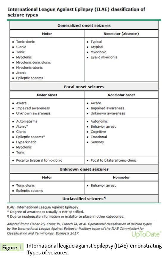
The ILAE Commission on Classification and Terminology proposed substantial changes to the 1989 classification system in 2010 and made further revisions in 2017 [5-7]. The current framework allows for diagnosis at four levels, depending on information and resources that are available, and also addresses the broad concepts of etiology and associated comorbidities at all levels (Figure 1).
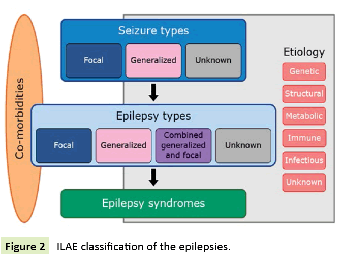
Idiopathic generalised epilepsy (IGE) is a common form of epilepsy, accounting for 20–40% of all epilepsies. IGE is clinically characterised by absence, myoclonic seizures, and tonic–clonic seizures with the electroencephalographic (EEG) pattern of bilateral, synchronous, and symmetrical spike and wave or polyspike and wave discharges. On the basis of the predominant seizure type and age of onset, the international classification recognises four main IGE sub syndromes: childhood absence epilepsy (CAE), juvenile absence epilepsy (JAE), juvenile myoclonic epilepsy (JME), and IGE with tonic–clonic seizures alone (GTCA) The term “idiopathic” is misleading, because it usually means “cause unknown,” which is not true here (Figure 2). Adult onset occurred in 13% of our patients with IGE; this supports the existence of a clinically distinct syndrome, which is consistent with reports of other investigators.
Brain AVMs occur in about 0.1 percent of the population, onetenth the incidence of intracranial aneurysms. Brain AVMs are considered sporadic congenital developmental vascular lesions, but their pathogenesis is not well understood. Rare cases of familial brain AVMs have been reported but it is unclear if these are coincidental or indicate true familial occurrence [8]. However, genetic variation may influence brain AVM development and clinical course [9,10].
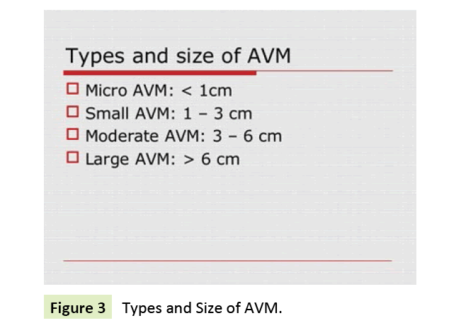
The angioarchitecture of brain AVMs is direct arterial to venous connections without an intervening capillary network. Both the arterial supply as well as the venous drainage may be by single or multiple vessels. Gliotic brain is usually admixed with the vascular tangle, and calcification may be seen in the vascular nidus and surrounding brain. The high-flow arteriovenous communication potentiates a variety of flow-related phenomena such as the development of afferent and efferent pedicle aneurysms, which occur in 20 to 25 percent of patients, and arterialization of the venous limb. Aneurysms can be a source of bleeding in patients with brain AVMs and are thought to worsen their prognosis. Abnormal flow and a vascular steal phenomenon have been suggested to underlie some clinical symptoms associated with brain AVMs (Figure 3).
Magnetic resonance angiography (MRA) is a group of techniques based on magnetic resonance imaging (MRI) to image blood vessels. Magnetic resonance angiography is used to generate images of the arteries in order to evaluate them for stenosis (abnormal narrowing), occlusion or aneurysms (vessel wall dilatations, at risk of rupture) [7]. MRA is often used to evaluate the arteries of the neck and brain, the thoracic and abdominal aorta, the renal arteries, and the legs (called a "run-off"). MRA sensitivity and specificity with arterial aneurysm it was 97% and 82%, also it was 97% and 82%, with stenosis of more than 50%, it was 98% and 46%, with stenosis of less than 50% and lastly it was 96% and 84% [8]. In the diagnosis of cerebral AVM nidus size S1: <3 and S2: 3-6 cm sensitivity was 100% but accuracy was 100% and 73.3% respectively.
DSA was 100% sensitive in the diagnosis of superficial and deep venous drainage AVM. Regarding the eloquence of brain area 15 had no eloquence by both MRA and DSA and identification of eloquence of brain area sensitivity was 73.3% and accuracy was 86.7%. The main feeding vessels was found (22, 73.3%) in both DSA and MRA findings. Distal vessels was seen (8, 26.7%) in DSA but not seen in MRA findings. Intranidal aneurysm and Angiopathic AVM were seen in 3(10.0%) and 4(13.3%) respectively in DSA. This study was carried out to diagnose the patients presented with cerebral AVM by MRA and DSA. MRA could not be evaluated flow status of AVM, distal feeding arteries, intranidal aneurysm and angiopathic AVM which could be detected by DSA. So, DSA is superior to MRA in diagnosis of cerebral AVM.
Objectives
Primary
Study prevalence of vascular abnormalities along patients with adult onset Idiopathic generalized Tonic clonic seizures.
Secondary
To revise the diagnostic criteria of adult onset Idiopathic generalized Tonic clonic seizures.
Materials and Methods
Research design: Prospective study.
Study population: 20 cases of IGTC seizures.
Inclusion criteria
• Age: >16 years old.
• Sex: NO sex difference.
• Family history of seizures: with or without family history of seizures.
• Normal MRI brain or CT brain, normal MRA.
• EEG: Positive/Negative EEG findings since SEIZURES are clinical diagnosis.
• Laboratory investigations: Normal metabolic profile (Na, K, Ca, Mg, Cl, kidney function, liver function).
• Patient had Adult onset Generalized tonic clonic seizures.
Exclusion criteria
• All cases of Secondary seizures and not presented by GTC seizures, below the age of 16 years old.
• Study settings: It is interventional clinical study.
• Duration of study: The proposed study will be conducted over a period of 6 months.
• Study instrument and validation procedure tools:
1) Clinical examination
2) Full Lab. Investigations
3) EEG
4) MRI brain, MRA brain
5) Diagnostic Digital subtraction angiography (DSA).
Validation procedure: Correlation between clinical examination finding, MRA brain, findings with DSA (All will validate each other).
Ethical issues: A written consent to be obtained from recruited subjects.
Methodology: Twenty outpatients fulfilling inclusion criteria of Idiopathic Generalized seizure (IGS) will be neurologically examined. MRI and MRA brain will be done to the patients followed by DSA. Data will be collected and analysed.
Result
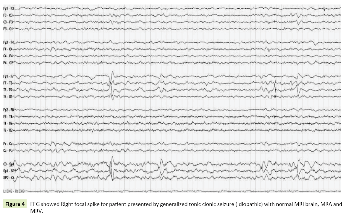
Demographic data: Age the youngest case was 17 years old and eldest was 28 years old. As regard types of generalized seizures, ten (50%) cases were generalized tonic clonic seizures, 6 (30%) cases were myoclonic seizures, 4 (20%) cases were absence seizures. For the twenty cases 16 cases (80%) showed generalized poly spikes and slow waves seen along all leads (Paroxysm of spikes and slow waves) on the other hand EEG showed unilateral persistent spikes for 4 cases (20%) of cases apart, corresponding MRI brain, MRA and MRV are completely normal in all cases. As regard DSA showed micro AVMs in 4 CASES (20%) cases (3 superficial right temporal, one superficial left parietal) (Figure 4).
Figure 4: EEG showed Right focal spike for patient presented by generalized tonic clonic seizure (Idiopathic) with normal MRI brain, MRA and MRV.
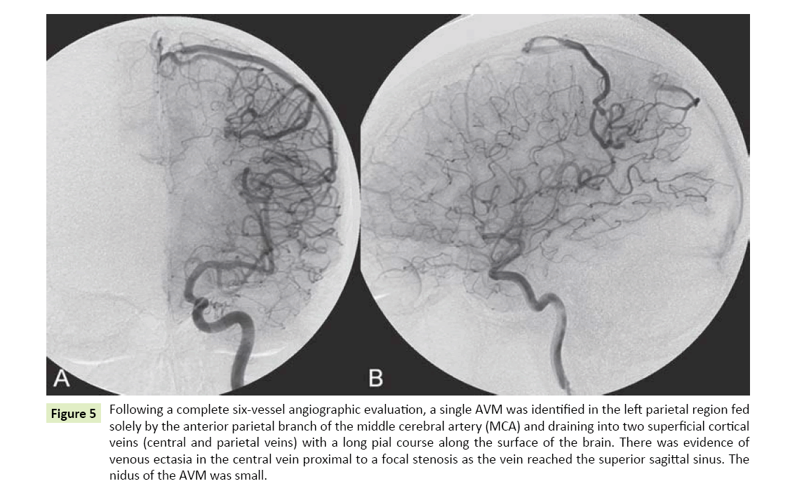
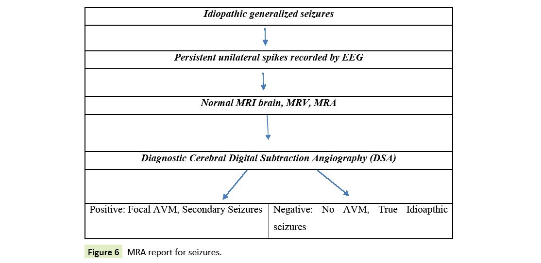
Correlation between DSA findings and EEG findings, EEG spikes were seen at same side of corresponding Micro AVMS recorded by DSA (Figures 5 and 6).
Figure 5: Following a complete six-vessel angiographic evaluation, a single AVM was identified in the left parietal region fed solely by the anterior parietal branch of the middle cerebral artery (MCA) and draining into two superficial cortical veins (central and parietal veins) with a long pial course along the surface of the brain. There was evidence of venous ectasia in the central vein proximal to a focal stenosis as the vein reached the superior sagittal sinus. The nidus of the AVM was small.
Discussion
Idiopathic generalized epilepsy (IGE) is a group of epileptic disorders that are believed to have a strong underlying genetic basis. Patients with an IGE subtype are typically otherwise normal and have no structural brain abnormalities. People also often have a family history of epilepsy and seem to have a genetically predisposed risk of seizures. IGE tends to manifest itself between early childhood and adolescence although it can be eventually diagnosed later. The genetic cause of some IGE types is known, though inheritance does not always follow a simple monogenic mechanism.
Brain AVMs occur in about 0.1 percent of the population, onetenth the incidence of intracranial aneurysms [1]. Supratentorial lesions account for 90 percent of brain AVMs; the remainder are in the posterior fossa. They usually occur as single lesions, but as many as 9 percent are multiple [10].
Brain AVMs underlie 1 to 2 percent of all strokes, 3 percent of strokes in young adults, and 9 percent of subarachnoid haemorrhages [11,12].
In our study we found that Prevalence of small AVM using DSA among patients with apparently Idiopathic Generalised seizures with persistent EEG focal spikes with secondary polyspike and normal MRI brain MRA, MRV is 20%. This is nearby study done by Garcine et al. on 2012 who found that Eleven to 33 percent of Patients with cortically-located, multiple, and superficial-draining AVMs are more likely to present with seizures [12]. Seizures are typically focal, either simple or partial complex, but often have secondary generalization.
So the question can we consider all cases of idiopathic generalized seizure with persistent focal spikes are really true idiopathic or they could be focal onset with very fast spread that the patient can’t recognize its focal onset clinically and based upon limited sensitivity of MRA BRAIN to detect very small AVM as mentioned before, DSA could help to exclude small sized AVMs.
Limitations
Our study is limited by the relatively small sample size.
Recommendations
As regard future Protocol for Cases with Clinical picture of generalized idiopathic seizures and persistent unilateral spikes recorded, DSA could be helpful to exclude true Idioathic from Secondary focal seizures with very fast spread to generalized seizures.
References
- Janz D, Beck-Mannagetta G, Sander T (1992) Do idiopathic generalised epilepsies share a common susceptibility gene? Neurology 42: 48-55
- King MA, Newton MR, Jackson GD, Fitt G, Silvapulle M, et al. (1998) Epileptology of the first-seizure presentation: a clinical, electroencephalographic, and magnetic resonance imaging study of 300 consecutive patients. Lancet 352: 1007-1011.
- Commission Report (1989) Commission on classification and terminology of the International League Against Epilepsy. Proposal for revised classification of epilepsies and epileptic syndromes. Epilepsiy 30: 389-399.
- Andermann F, Berkovic SF (2001) Idiopathic generalised epilepsy with generalised and ther seizures in adolescence. Epilepsia 42: 317-320.
- Gilliam F, Steinhoff BJ, Bittermann HJ (2000) Adult myoclonic epilepsy: a distinct syndrome of idiopathic generalized epilepsy. Neurology55: 1030-1033.
- Panayiotopoulos CP, Koutroumanidis M, Giannakodimos S (1997) Idiopathic generalised epilepsy in adults manifested by phantom absences, generalised tonic-clonic seizures, and frequent absence status. J Neurol Neurosurg Psychiatry63: 622-627.
- Cukur T, Lee JH, Bangerter NK, Hargreaves BA, Nishimura DG (2009). "Non-contrast-Enhanced Flow-Independent Peripheral MR Angiography with Balanced SSFP". Magnetic Resonance in Medicine 61 : 1533-1539.
- Chowdhury AH, Ghose SK, Mohammad QD, Habib M, Khan SU, et al. (2015) “Digital Subtraction Angiography is Superior to Magnetic Resonance Angiography in Diagnosis of Cerebral Arteriovenous Malformation.” Mymensingh Med J 24: 356-365.
- Fisher RS, Acevedo C, Arzimanoglou A, Bogacz A, Cross JH, et al. (2014)“ILAE official report: a practical clinical definition of epilepsy.” Epilepsia 55: 475-482.
- Beijnum VJ, Worp V, Schippers HM, Nieuwenhuizen OV, Kappelle LJ, et al. (2007) “Familial occurrence of brain arteriovenous malformations: a systematic review.” J Neurol Neurosurg Psychiatry 78: 1213-1217.
- Hashimoto T, Lawton MT, Wen G, Yang GY, Chaly T, et al. (2004) “Gene microarray analysis of human brain arteriovenous malformations.” Neurosurgery 54: 410-423; 423-425.
- Garcin B, Houdart E, Porcher R, Manchon E, Bresson D, et al. (2012) “Epileptic seizures at initial presentation in patients with brain arteriovenous malformation.” Neurology 78: 626-631.
Open Access Journals
- Aquaculture & Veterinary Science
- Chemistry & Chemical Sciences
- Clinical Sciences
- Engineering
- General Science
- Genetics & Molecular Biology
- Health Care & Nursing
- Immunology & Microbiology
- Materials Science
- Mathematics & Physics
- Medical Sciences
- Neurology & Psychiatry
- Oncology & Cancer Science
- Pharmaceutical Sciences
