ISSN : 2471-8548
Journal of Neuropsychiatry
From molecular to behavior: Higher Order occipital cortex in major depressive disorder
European Conference on Psychiatry
July 25, 2022 | Webinar
Xue Mei Song
Zhejiang University School of Medicine, 310029, China
ScientificTracks Abstracts: J Neuropsychiatry
Abstract
Statement of the Problem: Medial prefrontal cortex and other regions like the occipital cortex (OC) exhibit abnormal neural activity in major depressive disorder (MDD). Their relationship to specific biochemical, psychophysical, and psychopathological changes remains unclear, though. For that purpose, we focus on a particular subregion in OC, namely middle temporal visual area (MT) that is known to mediate the perception of visual motion. Methodology & Theoretical Orientation: Using Ultra-high-field 7T MRI, including resting state fMRI and proton magnetic resonance spectroscopy (MRS), the amplitude of low-frequency fluctuations (ALFF) of the blood oxygen level-dependent signal in MT, MT-seeded functional connectivity, and GABA in MT were investigated. Applying the vision motion psychophysical task, the motion suppression index of subjects was also examined. Findings: We find significantly elevated neural variability (as measured by ALFF) in MT together with decreases in both MT GABA and motion suppression in our MDD sample. Unlike in healthy subjects, MT neural variability no longer modulates the relationship of MT GABA and motion suppression in MDD. MT also exhibits reduction in global inter-regional functional connectivity to medial prefrontal cortex in MDD. Finally, elevated MT ALFF relates to specifically retardation in behavior as measured by the Hamilton subscore. Conclusion & Significance: Our findings demonstrate the importance of higher order OC, that is, MT for local-regional function, global topography, and specific depressive symptoms in acute MDD. That singles out MT as a strong candidate biomarker and potential treatment target in MDD.
Biography
Xue Mei Song has her expertise in studying the visual perception and its neural mechanism for more than 20 years. From 2018, she used a cross-species visual stimulus-inter and surround interaction, and combined the technology of ultra-high-field (7T) MRI, to study the neural mechanism of MDD. She and collaborates firstly found that the MDD patients suffer from perceptual changes in following visual speed which is related to the brain’s biochemical (Molecular Psychiatry, 2021). Later study found the dysfunction in middle temporal visual area (MT) of MDD, from molecular to neural, brain functional connectivity, behavior, and psychopathological levels (Cerebral Cortex, 2022).
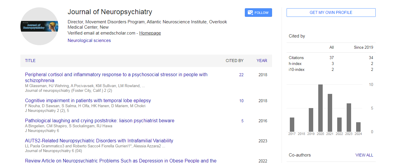
Google Scholar citation report
Citations : 37
Journal of Neuropsychiatry received 37 citations as per Google Scholar report
Abstracted/Indexed in
- Google Scholar
- China National Knowledge Infrastructure (CNKI)
- Secret Search Engine Labs
- Euro Pub
Open Access Journals
- Aquaculture & Veterinary Science
- Chemistry & Chemical Sciences
- Clinical Sciences
- Engineering
- General Science
- Genetics & Molecular Biology
- Health Care & Nursing
- Immunology & Microbiology
- Materials Science
- Mathematics & Physics
- Medical Sciences
- Neurology & Psychiatry
- Oncology & Cancer Science
- Pharmaceutical Sciences