ISSN : 2574-2825
Journal of Nursing and Health Studies
Fluid removal in peritoneal dialysis
27th Edition of World Congress on Nursing Education & Research
April 23-25, 2018 Rome, Italy
Raymond T Krediet
Academic Medical Centre, Netherlands
Posters & Accepted Abstracts: J Nurs Health Stud
DOI: 10.21767/2574-2825-C1-003
Abstract
Removal of uremic waste products and excess of accumulated body fluid are the goals of peritoneal dialysis (PD) in the treatment of patients with chronic renal failure. The peritoneum used as a dialysis membrane consists of the mesothelial layer and interstitial tissue, in which blood- and lymphatic vessels are present. The microvessels allow transport of solutes and water from the blood to the dialysis fluid in the peritoneal cavity. Solutes like urea are transported by diffusion across the vascular wall; fluid removal (ultrafiltration) requires a pressure gradient. The latter consists of the intravascular hydrostatic pressure, which drives fluid out of the microvessels to the interstitium through interendothelial pores, but also of an osmotic pressure gradient. The latter is created by adding high dosages of glucose to the dialysis fluid. This is only effective, because the transcellular water channel aquaporin-1 (AQP-1) is present in peritoneal endothelial cells. AQP-1 is permeable to water only, not to solutes like glucose and Na+. Consequently AQP-1 allows free water (water only) transport (FWT). The osmotic gradient contributes to SPFT to a limited extent only. During the first few years of PD about 40% of the ultrafiltered volume consists of FWT and 60% of small-pore fluid transport (SPFT). Loss of ultrafiltration capacity occurs especially in long-term PD patients. It is mostly associated with high solute transport rates, suggestive of an enlarged vascular surface area leading to rapid disappearance of the osmotic gradient. This affects especially SPFT. A longitudinal study showed a marked reduction of SPFT after 4 years of PD, possibly due to vascular abnormalities. FWT is markedly decreased in patients who develop encapsulating peritoneal sclerosis. Binding of free water by a collagen increase is the most likely explanation. Determination of both SPFT and FWT are essential in the followup of PD patients. r.t.krediet@amc.uva.nl
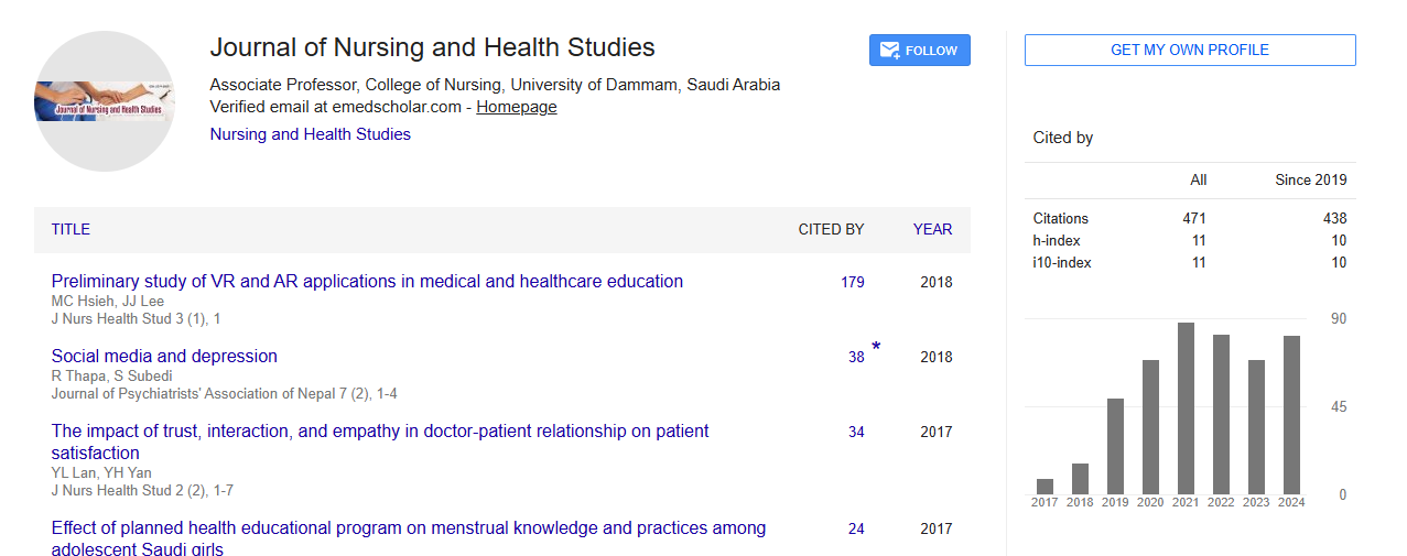
Google Scholar citation report
Citations : 471
Journal of Nursing and Health Studies received 471 citations as per Google Scholar report
Abstracted/Indexed in
- Google Scholar
- Open J Gate
- China National Knowledge Infrastructure (CNKI)
- Cosmos IF
- Secret Search Engine Labs
- Euro Pub
Open Access Journals
- Aquaculture & Veterinary Science
- Chemistry & Chemical Sciences
- Clinical Sciences
- Engineering
- General Science
- Genetics & Molecular Biology
- Health Care & Nursing
- Immunology & Microbiology
- Materials Science
- Mathematics & Physics
- Medical Sciences
- Neurology & Psychiatry
- Oncology & Cancer Science
- Pharmaceutical Sciences