ISSN : ISSN No. 2472-1123
Journal of Organic & Inorganic Chemistry
Cancer cell imaging using in situ generated gold nanoclusters
8th Edition of International Conference on Chemistry Education and Research
August 27-28, 2018 Zurich, Swit zerland
Md Asif Amin and Kankan Bhattacharyya
Balurghat Mahila Mahavidyalaya, India Indian Institute of Science Education and Research, India
Posters & Accepted Abstracts: J Org Inorg Chem
DOI: 10.21767/2472-1123-C5-015
Abstract
In situ generated fluorescent gold nanoclusters (Au-NCs) are used for bio-imaging of three human cancer cells, namely, lung (A549), breast (MCF7), and colon (HCT116), by confocal microscopy. The amount of Au-NCs in non-cancer cells (WI38 and MCF10A) is 20–40 times less than those in the corresponding cancer cells. The presence of a larger amount of glutathione (GSH) capped Au-NCs in the cancer cell are ascribed to a higher glutathione level in cancer cells. The Au-NCs exhibit fluorescence maxima at 490–530 nm inside the cancer cells. The fluorescence maxima and matrix-assisted laser desorption ionization (MALDI) mass spectrometry suggest that the fluorescent Au-NCs consist of GSH capped clusters with a core structure (Au8-13). Timeresolved confocal microscopy indicates a nanosecond (1–3 ns) lifetime of the Au-NCs inside the cells. This rule out the formation of aggregated Au–thiolate complexes, which typically exhibit microsecond (.1000 ns) lifetimes. Fluorescence correlation spectroscopy (FCS) in live cells indicates that the size of the Au-NCs is .1–2 nm. For in situ generation, we used a conjugate consisting of a room-temperature ionic liquid (RTIL, [pmim][Br]) and HAuCl4. Cytotoxicity studies indicate that the conjugate, [pmim][AuCl4], is non-toxic for both cancer and non-cancer cells.
Biography
E-mail:
mdasifamin007@gmail.com
Google Scholar citation report
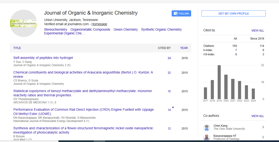
Citations : 150
Journal of Organic & Inorganic Chemistry received 150 citations as per Google Scholar report
Abstracted/Indexed in
- Google Scholar
- China National Knowledge Infrastructure (CNKI)
- Directory of Research Journal Indexing (DRJI)
- WorldCat
- Geneva Foundation for Medical Education and Research
- Secret Search Engine Labs
Open Access Journals
- Aquaculture & Veterinary Science
- Chemistry & Chemical Sciences
- Clinical Sciences
- Engineering
- General Science
- Genetics & Molecular Biology
- Health Care & Nursing
- Immunology & Microbiology
- Materials Science
- Mathematics & Physics
- Medical Sciences
- Neurology & Psychiatry
- Oncology & Cancer Science
- Pharmaceutical Sciences