ISSN : 2471-8041
Medical Case Reports
A rare case report of complete expression of pentalogy of cantrell: from radiology perspective Antenatal ultrasound, fetal echo and fetal MRI, post termination radiography and 3D CT findings with a clinical autopsy correlation:
8th Edition of International Conference on Clinical and Medical Case Reports
May 28-29, 2018 London, UK
Leul Adane, Alemayehu Bedane, Fitehanegest Tefera and Satyasai Panda
Saint Paul Hospital Millennium Medical College, Ethiopia
Posters & Accepted Abstracts: Med Case Rep
DOI: 10.21767/2471-8041-C1-003
Abstract
Pentalogy of cantrell consists of an extensive defect of the thoraco-abdominal wall, which has nearly always a lethal prognosis. The defect is characterized by the association of five anomalies: omphalocele, cardiac ectopia, absence of the distal portion of the sternum, absence of the anterior diaphragm and absence of the parietal diaphragmatic pericardium. It has a rare frequency of about 5.5 per 1,000,000 live births. There is a common association with intra cardiac anomalies such as ventricular septum defect, tetralogy of fallot and transposition of great vessels. The pathogenesis remains unclear. Here we present an imaging findings with antenatal two dimensional (2D) and three dimensional (3D) ultrasound and fetal magnetic resonance imaging (MRI) in a 20 weeks of gestation with a multiple anomalies, based on which the diagnosis of complete pentalogy of cantrell was given with a brief literature. Post mortem radiography, 3D computed tomography (CT) and clinical autopsy were performed additionally to enhance the visualization of fetal anomalies and to confirm the diagnosis. Extensive imaging of cardiac, thoracic and abdominal malformations by ultrasound and MRI is complementary for a clear diagnosis and counseling of the patient.
Biography
E-mail: leuladane2@gmail.com
Google Scholar citation report
Citations : 241
Medical Case Reports received 241 citations as per Google Scholar report
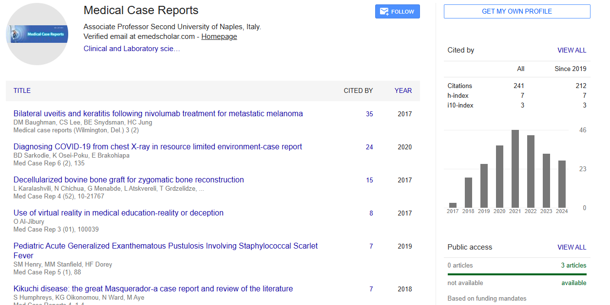
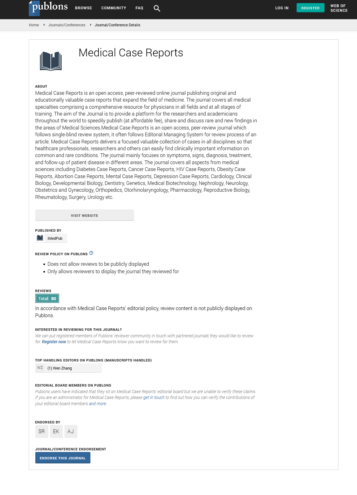
Medical Case Reports peer review process verified at publons
Abstracted/Indexed in
- Google Scholar
- China National Knowledge Infrastructure (CNKI)
- Cosmos IF
- Directory of Research Journal Indexing (DRJI)
- WorldCat
- Publons
- Secret Search Engine Labs
- Euro Pub
Open Access Journals
- Aquaculture & Veterinary Science
- Chemistry & Chemical Sciences
- Clinical Sciences
- Engineering
- General Science
- Genetics & Molecular Biology
- Health Care & Nursing
- Immunology & Microbiology
- Materials Science
- Mathematics & Physics
- Medical Sciences
- Neurology & Psychiatry
- Oncology & Cancer Science
- Pharmaceutical Sciences