ISSN : 2634-7806
Journal of Clinical and Molecular Pathology : Open Access
Intravascular Synovial Sarcoma of the Femoral Vein: A Case Report
Gaurav Singal1, Anshoo Agarwal2 and Hala Abdel Dayem Mouhamed3*
1Consultant, Ivy Hospital, Mohali Chandigarh, India
2Deptartment of Pathology, Faculty of Medicine, Northern Borders University, Kingdom Saudi Arabia
3Deptartment of Pathology, Faculty of Medicine, Suez Canal University, Ismailia, Egypt
- *Corresponding Author:
- Hala Abdel-Dayem Mouhamed
Department of Pathology, Faculty of Medicine
Suez Canal University, Egypt
Tel: 00966509091425
E-mail: hala.abdeldayem@yahoo.com
Received date: March 06, 2017; Accepted date: April 12, 2017; Published date: April 18, 2017
Citation: Singal G, Agarwal A, Mouhamed D. Intravascular Synovial Sarcoma of the Femoral Vein: A Case Report. J Clin Mol Pathol 1: 11.
Copyright: © 2017 Mouhamed D et al. This is an open-access article distributed under the terms of the Creative Commons Attribution License, which permits unrestricted use, distribution, and reproduction in any medium, provided the original author and source are credited.
Abstract
A case of intravascular biphasic synovial sarcoma arising from the wall of the femoral vein in a -41-year old man is being reported. The patient presented with swelling and localized right leg pain. The diagnosis was made intraoperative. The tumor was resected and vascular reconstruction was performed. For post-operative radiation therapy & chemotherapy, the patient was referred to the oncologist. This is the very rare case of an intravascular synovial sarcoma known to be reported in the medical literature. Few previous cases arose from highlights about the characteristic clinical pattern and the location in large veins of the lower extremity and trunk. Primary synovial sarcoma of a blood vessel is extremely rare, and this report shares the clinical presentation and treatment of this patient. Synovial sarcoma must be considered in the differential diagnosis of intravascular tumors.
Keywords
Femoral vein; Synovial sarcoma; Vascular tumor
Introduction
Primary vascular tumors are rare entities; sarcomas are the most common type [1-3]. A few hundred cases have been reported since Perl’s first description in 1871 [4]. Synovial sarcoma is a well-defined entity with characteristic clinical and morphological features. It occurs predominantly in adolescents and young adults, and arises most commonly from para-articular soft tissues, particularly around the knee. We report a case of synovial sarcoma arising from the wall of the femoral vein in a 41-year-old man.
Case Report
A 41 years old male presented with the complaint of swelling on the right upper thigh which he noticed 2 weeks ago. The swelling was associated with localized pain. There was no history of peripheral edema. The initial radiological evaluation was suggestive of tumor arising in the vicinity of blood vessels. Doppler venous scan done for the status of deep veins was suggestive of chronic DVT of the popliteal and proximal superficial veins. Further evaluation with MRI indicated the possibility of synovial sarcoma. There were no palpable lymph nodes and no evidence of a tumor in any other part of the limb or elsewhere.
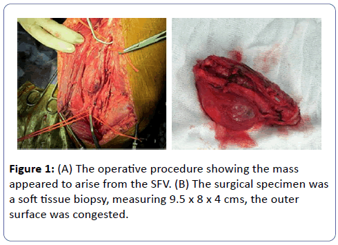
During surgery, the mass was found to be arising from the superficial femoral vein (SFV) just where it drains into the common femoral vein (CFV) involving almost 10 cms of the venous segment (Figures 1A and B).
It was adherent to the femoral artery which was excised along with vein for wider surgical clearance. The femoral artery was reconstructed with expanded Polytetrafluoroethylene (ePTFE) graft. SFV was not reconstructed because of the presence of chronic changes.
The postoperative course was uneventful. The patient is doing well and is referred to the medical oncologist for chemotherapy/ radiation therapy since these tumors are known to metastasize to the lung and the liver.
Pathological Findings
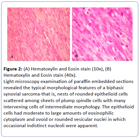
The surgical specimen was a soft tissue biopsy, measuring 9.5 x 8 x 4 cms, the outer surface was congested. On cutting tumor, it was fleshy homogenous with foci of hemorrhage. On serial section, a thick-walled vessel was identified (Figure 1). Light microscopy examination of paraffin wax embedded sections showed tumor composed predominantly of spindle cells disposed of in interlacing fascicles and sheets. The tumor cells had oblong blunt end nucleus and abundant pink cytoplasm (Figures 2A and B).
Figure 2: (A) Hematoxylin and Eosin stain (10x), (B) Hematoxylin and Eosin stain (40x). Light microscopy examination of paraffin embedded sections revealed the typical morphological features of a biphasic synovial sarcoma-that is, nests of rounded epithelioid cells scattered among sheets of plump spindle cells with many intervening cells of intermediate morphology. The epithelioid cells had moderate to large amounts of eosinophilic cytoplasm and ovoid or rounded vesicular nuclei in which occasional indistinct nucleoli were apparent.
Some areas showed mitotic activity 5-6/10HPF. Areas of hemorrhage were seen without any necrosis. Diagnosis made was a malignant mesenchymal tumor with a possibility of synovial sarcoma.
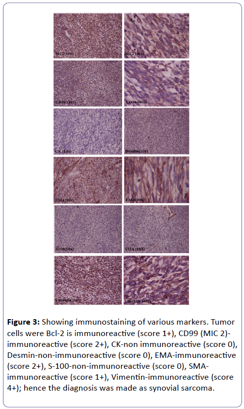
Immunohistochemistry of (Vimentin, Cytokeratin, Smooth Muscle actin, Desmin, CD-99, Epithelial Membrane Antigen, S-100, Bcl-2) was performed to confirm the diagnosis. Immunohistochemistry revealed the following results that showed in (Figure 3): Vimentin-immunoreactive (score 4+) in tumor cells, Cytokeratin-non-immunoreactive (score 0) in tumor cells, Smooth Muscle actin-Immunoreactive (score 1+) in tumor cells, Desmin-non-immunoreactive (score 0) in tumor cells, CD99 (MIC 2)-immunoreactive (score 2+) in tumor cells, Epithelial Membrane Antigen-immunoreactive (score 2+) in tumor cells, S-100-non-immunoreactive (score 0) in tumor cells, Bcl-2 is immunoreactive (score 1+). Since Bcl-2 had immunoreactive (score 1+) hence the diagnosis was made as synovial sarcoma (Figure 3).
Figure 3: Showing immunostaining of various markers. Tumor cells were Bcl-2 is immunoreactive (score 1+), CD99 (MIC 2)- immunoreactive (score 2+), CK-non immunoreactive (score 0), Desmin-non-immunoreactive (score 0), EMA-immunoreactive (score 2+), S-100-non-immunoreactive (score 0), SMAimmunoreactive (score 1+), Vimentin-immunoreactive (score 4+); hence the diagnosis was made as synovial sarcoma.
Discussion
Synovial sarcoma commonly arises from the para-articular soft tissues of the extremities. It is most prevalent in adolescents and young adults (15 to 40 years old), and there is a slight male predominance (1.2:1). It usually arises in close association with tendon sheaths, bursae, and joint capsules, but is uncommonly encountered within joint cavities [5].
Rarely it is found at sites having no apparent synovial structures, such as the tongue, para-pharyngeal region, and abdominal wall, and consequently, its origin from preformed synovial tissues is doubtful. Our case and those previously reported [6-8], of a biphasic synovial sarcoma arising from, and entirely contained by, a blood vessel appears to exclude a synovial origin for these tumors. Robertson et al. reported that although the number of cases reported is very small, a characteristic clinical pattern seems to be emerging that is location in large veins of the lower extremity and trunk in young adult women [9].
Ultrasound with a high-frequency probe associated with color Doppler may provide the diagnosis when the tumor is intraluminal. Many authors consider MRI to be the examination of choice for evaluating soft tissue mass of undetermined diagnosis and structural content. It provides information about tumor localization, size, and intraluminal and extra luminal involvement8. Appropriate interpretation and diagnosis are crucial for effective management. Doppler ultrasound and MRI are useful to establish early diagnosis at the non-tumoral stage. Improvement in the prognosis of synovial sarcoma may justify perioperative chemotherapy before and after radical surgical excision [10].
Histologic Characteristics
Microscopically, the tumor is made up of epithelial and sarcomatous components. The sarcomatous structure can lead to an incorrect diagnosis of a malignant schwannoma or fibrosarcoma. Immunohistochemistry staining is the key to a definitive diagnosis. The monophasic form exhibits primarily sarcomatous features; the monophasic epithelial form is rare.
Treatment
The relatively small number of patients with synovial sarcoma of the blood vessels precludes us from drawing firm conclusions with respect to a precise treatment protocol. The primary mode of treatment is wide surgical excision includes a normalappearing tissue cuff to obtain tumor-negative margins. Multimodality treatment results appear to be better than those seen with single-modality treatment or salvage therapy. The role of radiotherapy is to reduce the risk of local recurrence following resection; radiation does not improve long-term survival. Chemotherapy also used to decrease the risk of distant metastasis [10].
Synovial sarcoma must be considered in the differential diagnosis of intravascular "tumors". Non-neoplastic and neoplastic conditions that may present in this fashion are very diverse and include intravascular fasciitis, pyogenic granuloma, epithelioid haemangioendothelioma, and leiomyosarcoma. Biphasic synovial sarcoma can be differentiated from these by virtue of its biphasic growth pattern with gland-like and spindled components, both of which show at least focal cytokeratin positivity [11].
This report also describes a removal of a synovial sarcoma arising from femoral vein necessitating removal of the femoral venous and arterial circulations. Femoral vein synovial sarcoma is especially challenging to manage. Successful outcome is predicated on revascularization with autologous vein and on a multidisciplinary approach using various soft tissue coverage strategies and wound management adjuncts.
Conclusions
Clinical presentation depends on the extra luminal/ intraluminal growth of the tumor mass in the vessels. If extra luminal it can result in nerve compression causing pain and if intraluminal it can mimic the symptoms of DVT. In our case, it was surprising that the patient never had any symptoms of DVT like limb edema etc. Management of such cases is challenging because of very low incidence with most of the data available in literature being single reports. We emphasize that synovial sarcoma also must be considered in the differential diagnosis of intravascular "tumors”.
References
- Dzsinich C, Gloviczki P, van Heerden JA, Nagorney DM, Pairolero PC, et al. (1992) Primary venous synovial sarcoma: a rare but letahal disease. J Vasc Surg 15: 595-603.
- Burke AP, Virmani R (1993) Sarcomas of the great vessels. A clinicopathologic study. Cancer 71: 1761-1773.
- Robertson NJ, Halawa MH, Smith MEF (1998) Intravascular synovial sarcoma. J Clinic Pathol 2: 172-173.
- Perl L (1871) Ein fall von sarkom der vena cava inferior. Virchows Arch 53: 378-383.
- Enzinger FM, Weiss SW (1995) Soft tissue tumors. (3edn) St Louis: Mosby, pp: 757-786.
- Miettinen M, Santavirta S, Slatis P (1987) Intravascular synovial sarcoma. Hum Pathol 18: 1075-1057.
- Shaw GR, Lais CJ (1993) Fatal intravascular synovial sarcoma in a 31-year-old woman. Hum Pathol 24: 809-810.
- McKinney CD, Mills SE, Fechner RE (1992) Intraarticular synovial sarcoma. Amy Surg Pathol 16: 1017-1020.
- Robertson NJ, Halawa MH, Smith MEF (1998) Intravascular synovial sarcoma. Journal of Clinical Pathology 2 : 172-173.
- Steinau HU, Daigeler A, Langer S, Steinsträsser L, Hauser J, et al. (2010) Limb salvage in malignant tumors. Semin Plast Surg 1: 18-33.
- Razek AA, Huang BY (2011) Soft tissue tumors of the head and neck: imaging-based review of the WHO classification. Radiographics 31: 1923-1954.
Open Access Journals
- Aquaculture & Veterinary Science
- Chemistry & Chemical Sciences
- Clinical Sciences
- Engineering
- General Science
- Genetics & Molecular Biology
- Health Care & Nursing
- Immunology & Microbiology
- Materials Science
- Mathematics & Physics
- Medical Sciences
- Neurology & Psychiatry
- Oncology & Cancer Science
- Pharmaceutical Sciences


