ISSN : ISSN: 2576-1455
Journal of Heart and Cardiovascular Research
Infective Endocarditis and Acute Myocardial Infarction
Rodolfo Pino*, Faro Manzella, Giuseppa Sciortino and Giovanni Polizzi
U.O. of Cardiology and U.T.I.C. Civic Hospital of Partinico, A.S.P. Palermo Via Circonvallazione n.1, Partinico (PA), Italy
- *Corresponding Author:
- Dr. Rodolfo Pino
U.O. of Cardiology and U.T.I.C. Civic Hospital of Partinico
A.S.P. Palermo Via Circonvallazione n.1
90047 Partinico (PA), Italy
Tel: +39-91-8911259
E-Mail: rodoraffa@tiscali.it
Received Date: June 09, 2017; Accepted Date: July 14, 2017; Published Date: July 21, 2017
Citation: Pino R, Manzella F, Sciortino G, Polizzi G (2017) Infective Endocarditis and Acute Myocardial Infarction. J Heart Cardiovasc Res. Vol. 1 No. 2:5
Abstract
Clinical Image
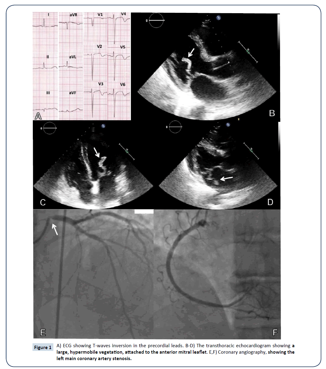
A 77-year-old male, with Parkinson disease and diabetes, was referred to the Intensive Cardiac Care Unit for a Non-ST-Elevation acute Myocardial Infarction; Troponin I levels peaked at 15395 pg/ml (normal <3 pg/ml) and the ECG showed a T-waves inversion in the precordial leads (Figure 1A).
The transthoracic echocardiogram showed left ventricular wall motion abnormalities of the apical septal and mid anterior septal segments, but also a large, serpiginous vegetation (30 × 5 mm), attached to the anterior mitral leaflet; it was hypermobile, with an impressive oscillation between left ventricle, in diastole (Figures 1B and 1C, arrows; in parasternal long-axis and apical four-chamber views, respectively) and left atrium in systole (Figure 1D, arrow); moreover, a moderate valvular regurgitation was present. A more detailed interview revealed that, the year prior to admission, the patient had several, recurrent bouts of fever, following a prostatic trans-urethral resection.
Blood cultures were positive for Enterococcus faecalis, and the patient was treated with ampicillin-sulbactam and vancomycin for 6 weeks; his condition progressively improved, but the vegetation’s size remained unchanged.
Despite the initial hypothesis of coronary septic embolism, coronary angiography (Figures 1E and 1F) showed a critical stenosis of the left main coronary artery (arrow).
The patient underwent surgical excision of the vegetation and mitral valve repair; in addition, a single saphenous vein bypass graft to the left anterior descending coronary artery, was performed. The microbiological analysis of the surgical specimen, confirmed the blood cultures’ results. The patient recovered well and postoperative period was uneventful [1,2].
The case we report is an unusual coexistence of two cardiac diseases, associated with a high mortality risk: the left main coronary artery stenosis and infective endocarditis with a very large, hypermobile vegetation.
References
- Conley MJ, Ely RL, Kisslo J, Lee KL, McNeer JF (1978) The prognostic spectrum of left main stenosis. Circulation 57:947-952.
- Thuny F, Di Salvo G, Belliard O, Avierinos JF, Pergola V(2005) Risk of embolism and death in infective endocarditis: prognostic value of echocardiography. A prospective multicenter study. Circulation 112:69-75.
Open Access Journals
- Aquaculture & Veterinary Science
- Chemistry & Chemical Sciences
- Clinical Sciences
- Engineering
- General Science
- Genetics & Molecular Biology
- Health Care & Nursing
- Immunology & Microbiology
- Materials Science
- Mathematics & Physics
- Medical Sciences
- Neurology & Psychiatry
- Oncology & Cancer Science
- Pharmaceutical Sciences

