Car's Deformation as an Indicator for the Management of Lateral Compression Type I Pelvic Fracture: A Case Report of Two Adults and Review of Literature
Orthopaedic department-specialized team for the management of pelvic fractures, 401 General Army Hospital of Athens (GAHA), Greece
- *Corresponding Author:
- Antonios Papasotiriou
2nd Orthopaedic department, 401 General
Army Hospital of Athens, Avenue Mesogion
138 & Katehaki, 11525 Greece.
Tel: +30 2107494385
E-mail: antpapas@hotmail.com
Received date: June 01, 2018; Accepted date: June 09, 2018; Published date: June 14, 2018
Citation: Papasotiriou AN, Darmanis S (2018) Car's Deformation as an Indicator for the Management of Lateral Com-Pression Type I Pelvic Fracture: A Case Report of Two Adults and Review of Literature. J Emerg Trauma Care. Vol.3 No.1:5.
Abstract
Pelvic ring fracture (PRF) is a result of high energy trauma especially in young adults. The management of lateral compression type I (LCI) as a subtype of PRF remains controversial. Fracture's displacement, pain of injured, fracture mobilization under anesthesia (MUA) are factors that determine the management of these injuries. However, there are no reports stated on car's deformation after an accident as a factor of decision making on pelvic fracture's treatment. We present two cases of adults sustained a surgical intervention in pelvis following an LCI injury with a review of literature. Photos of car's deformation following the collision add extra information about the severity of pelvic fracture. In conclusion car's deformation could be used as an indicator for the management of these injuries in combination with the above mentioned factors.
Keywords
Lateral compression; Car deformation; Pelvic fracture
Introduction
Pelvic ring fractures represent nowadays the 3% to 8% of all skeletal injuries [1]. A classification system that gained a worldwide recognition is that of Young and Burgess [2]. According to this classification these injuries are categorized in 3 types according to the direction of force that acts on pelvic ring; anterior-posterior compression (APC), lateral compression (LC), and vertical shear (VS). There are 3 subtypes of APC and LC with LCI to represent a pelvic fracture combined with an impaction fracture to the sacrum [3,4].
The treatment of pelvic fracture lateral compression type I (LCI) remains controversial [5,6]. The need to conservative or operative management is unclear. Factors as fracture displacement and checking fracture stability with mobilization under anesthesia (MUA), patient’s ability of mobilization and pain of injured determine the decision making for the treatment of these fractures [7,8].
The objective of this study is to introduce the deformation of the vehicle after a road traffic collision (RTC) as a possible new factor that could determine the management of LCI injuries [9].
To support the above scope, we report 2 cases of pelvic fracture type LCI after an RTC surgically treated with a review of literature.
Case Presentation
Case 1
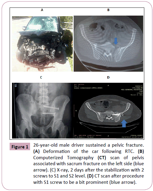
A 26-year-old male driver who was collided by another vehicle in April 2017. The crush happened on the side of the driver that was the left one. The driver was wearing his seat belt. He was admitted to our hospital (level I, trauma center) at the emergency trauma unit and was referred as a stable patient. After the necessary imaging evaluation (x-rays, (anteroposterior (AP) view of pelvis) and a computerized tomography (CT) scan of the pelvis, he was diagnosed with a pelvic fracture type LCI including the left sacrum fracture type Denis II, the left superior pubic ramus (in zone III according to Nakatami), the left ischiopubic ramus and the left transverse process of L5 lumbar spine [2,9,10]. No other injuries were reported. Following a clinical examination and examining the photos from the accident, the fracture to L5 process was diagnosed to be a result of a rotational and not of a vertical force. All the fractures were minimal displaced with the sacrum fracture to be a complete fracture with displacement between 1 and 2 cm in axial view (Figure 1).
Figure 1: 26-year-old male driver sustained a pelvic fracture.(A) Deformation of the car following RTC. (B) Computerized Tomography (CT) scan of pelvis associated with sacrum fracture on the left side (blue arrow). (C) X-ray, 2 days after the stabilization with 2 screws to S1 and S2 level. (D) CT scan after procedure with S1 screw to be a bit prominent (blue arrow).
Case 2
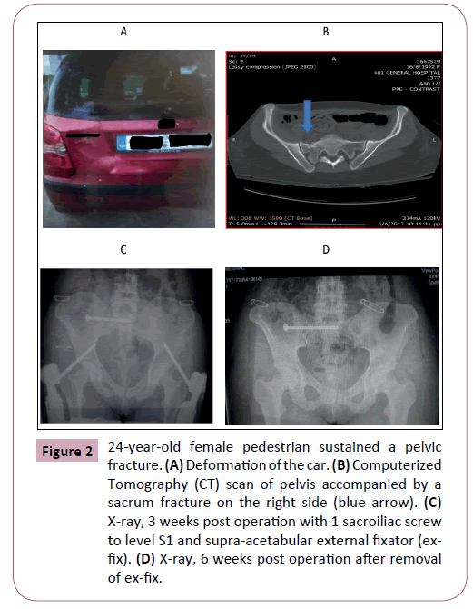
A 24-year-old female pedestrian who got hit by her own car in May 2017 which was left without handbrake and driver. That resulted in a lateral compression of her pelvis as she was found between the posterior left corner of her left-hand drive vehicle and a stone fence, in her attempt to stop the car. Initially, she was admitted to a provincial hospital (level III, trauma center) and was referred to us ten days after the initial admission. The patient was stable without other injuries than the pelvic fracture. Photos from the accident were also available to us, showing the dent of the vehicle on its left posterior corner to be approximately 30 × 30 cm with depth of 2 to 3cm. The appropriate imaging control (x-rays, CT) revealed a lateral compression pelvic fracture type LCI, including the right sacrum fracture type Denis II, the right ischiopubic ramus and the left pubic ramus (in zone II ) [2,9,10]. The sacrum fracture was complete with all the above fractures to be displaced less than 1 cm (Figure 2).
Figure 2: 24-year-old female pedestrian sustained a pelvic fracture. (A) Deformation of the car. (B) Computerized Tomography (CT) scan of pelvis accompanied by a sacrum fracture on the right side (blue arrow). (C) X-ray, 3 weeks post operation with 1 sacroiliac screw to level S1 and supra-acetabular external fixator (exfix). (D) X-ray, 6 weeks post operation after removal of ex-fix.
Result
Both these two young adults have been operated on 2 weeks after the initial injury. Although the male was admitted to our hospital immediately after the accident, he was referred with delay to a pelvic surgeon of the orthopaedic department. Up to the moment of the operation, both patients were unable to move out of bed as they were in severe pain localized at the back of the pelvis respectively at the site of the sacrum fracture, even though they were under analgesic therapy. The male underwent percutaneously stabilization in supine position with 2 iliosacral 7.3 cannulated screws with washer and short thread in S1 and S2 level respectively. The 2-dimensional (2D) fluoroscopy has been used. The pelvic fracture of the female was also operated on percutaneously with the patient to be in supine position. One iliosacral 7.3 cannulated screw with washer and short thread has been performed under 2D fluoroscopy to S1 level in combination with a supra-acetabular external fixator (ex-fix) using 1 pin 5.0 mm in each side of the pelvis [8-12]. The day after the operation both patients were able to mobilize using crutches with toe touch weight bearing at the site of the sacrum fracture for a period of three weeks. After that period partial weight bearing was allowed using always 2 crutches and almost in 10 weeks’ time they were able to walk without any support. About the injured female, the ex-fix has been removed in 6 weeks’ time, as there was confirmation of fracture healing in anterior pelvis using x-rays of pelvis (AP, inlet, outlet views). Both patients had excellent results in Majeed score in a 5 months’ post-operational period [1]. The healing of the fracture was confirmed with x-rays of the pelvis in 3 months after intervention. Regarding the female patient, a stone in bladder was found 3 months after the surgery which was removed under cystoscopy. She suffered painful urination and a CT scan of pelvis and ultra sound (U/S) of bladder was performed to investigate and finally diagnose the stone. Regarding the male, the S1 screw was a bit prominent without provoking any dysfunction for the injured. No other implications were reported to our patients (Figures 1 and 2).
Discussion
The management of LCI pelvic fractures remains a debate between the orthopedists [5,6]. Nowadays, in literature, not all the LCI pelvic fractures are the same. There are cases where the fracture to the sacrum is complete or not [8]. This fact in combination with the displacement of the anterior pelvis, determines the stability of the pelvic ring [7,8]. Support the idea that mobilization under anesthesia (MUA) could suggest the stability of the pelvis and consequently determine the decision for the management of the fracture [7,8]. In addition, the clinical evaluation of the patient is also a critical point for the final decision. Needless to say, that pain around pelvis and difficulties in mobilization must be taken under consideration [5].
In our study, both patients sustained a pelvic fracture with minimal displacement but with severe pain in posterior pelvis around the sacrum and were unable to mobilize themselves out of bed. So, it is assumed that the type of fracture alone is not an index of decision making, as does not indicate the severity of injury. This observation strengthens the idea of MUA of the pelvis [7,8]. In our study, the MUA hasn’t been introduced as it is probably considered a weak point of the manuscript. However, the information that concerns the stability of pelvis using the MUA could be also deduced through other studies or observations as the symptom of pain. It was observed through our 1st case that there was an ipsilateral fracture of the L5 transverse process caused probably by rotational force. This type of fracture could indicate instability of pelvis and therefore a surgical intervention should be performed [13]. However, an L5 transverse process fracture doesn’t merely predict instability of pelvis [14]. Finally, our decision for surgical intervention was supported by the imaging of pelvis, the clinical evaluation of the injured as well as the photos presented from the damage of both cars [15].
In our best knowledge, there is no study that indicates the vehicle deformation as an additional factor of surgical intervention in pelvic fracture type LCI. However, Stefanopoulos et al. after their study of 48 vehicle crashes in Greece, found that cars’ deformation is a significant factor affecting drivers’ and passengers’ injuries [16]. McCoy in his study in 1989 suggests the biomechanical parameters of pelvic injury after an RTC [17]. In addition, pelvic disruption is examined after a lateral impact [9,18].
Photos derived from the accident scene were available for both injuries. With a careful assessment of these photos, conclusions were drawn about the intensity of collision that provoked the pelvic fracture [19]. Even if the LCI pelvic fracture seems to be undisplaced the deformation of the vehicle could probably suggest the stability of LCI fracture, and therefore the decision making could also be supported by this information. So, more studies could elaborate on the topic of metals’ deformation after a crush, and correlate the deformation depicted from various accidents’ photos with the type and stability of pelvic fracture [9,16-19]. So, the subjective estimation on stability of a pelvic fracture following an RTC, could be realistic. So, gained information through photos of RTC could replace the MUA whose disadvantages include surgical time and potential risk of anesthesia for the injured.
Conclusion
The decision about the management of the pelvic fracture LCI, could be supported by one or more of the following factors; the clinical evaluation of the patient (i.e. mobilization or localized pain), the imaging evaluation of pelvis and the stability of the fracture checked by MUA and/or the deformation of the vehicle after the accident [4-9,16-18].
References
- Papasotiriou AN, Prevezas N, Krikonis K, Alexopoulos EC (2017) Recovery and return to work after a pelvic fracture. Saf Health Work 8: 162-168.
- Burgess AR, Eastridge BJ, Young JW, Elison TS, Elison PS, et al. (1990) Pelvic ring disruptions: Effective classification system and treatment protocols. J Trauma 30: 848-856.
- Lefaivre KA, Padalecki JR, Starr AJ (2009) What constitutes a young and burgess lateral compression-I (OTA 61-B2) pelvic ring disruption? A description of computed tomography-based fracture anatomy and associated injuries. J Orthop Trauma 23: 16-21.
- Hagen J, Castillo R, Dubina A, Gaski G, Manson TT, et al. (2016) Does surgical stabilization of lateral compression-type pelvic ring fractures decrease patients’ pain, reduce narcotic use, and improve mobilization? Clin Orthop Relat Res 474: 1422-1429.
- Lykomitros VA, Papavasiliou KA, Alzeer ZM, Sayegh FE, Kirkos JM, et al. (2010) Management of traumatic sacral fractures: A retrospective case-series study and review of the literature. Injury 41: 266-272.
- Kanakaris NK, Tzioupis C, Nikolaou VS, Giannoudis PV (2009) Lateral compression type I injuries of the pelvic ring stable? Injury Extra 40: 183-235.
- Saqi HC, Coniglione FM, Stanford JM (2011) Examination under anesthetic for occult pelvic instability. J Orthop Trauma 25: 529-536.
- Tosounidis T, Kanakaris N, Nikolaou V, Tan B, Giannoudis PV, et al. (2012) Assessment of lateral compression type 1 pelvic ring injuries by intraoperative manipulation: Which fracture pattern is unstable? Int Orthop 36: 2553-2558.
- Salzar RS, Genovese D, Bass CR, Bolton JR, Guillemot H, et al. (2009) Load path distribution within the pelvic structure under lateral loading. Inter J Crashworthiness 14: 99-110.
- Starr AJ, Nakatami T, Reinert CM, Cederberq K (2008) Superior pubic ramus fractures fixed with percutaneous screws: What predicts fixation failure? J Orthop Trauma 22: 81-87.
- Denis F, Davis S, Comfort T (1998) Sacral fractures: An important problem. Retrospective analysis of 236 cases. Clin Orthop Relat Res 227: 67–81.
- Bellabarba C, Ricci WM, Bolhofner BR (2006) Distraction external fixation in lateral compression pelvic fractures. J Orthop Trauma 20: 7-14.
- Starks I, Frost A, Wall P, Lim J (2011) Is a fracture of the transverse process of L5 a predictor of pelvic fracture instability? J Bone Joint Surg Br 93: 967-969.
- Maqungo S, Kimani M, Chhiba D, McCollum G, Roche S, et al. (2015) The L5 transverse process fracture revisited: Does its presence predict the pelvis fracture instability? Injury 46: 1629-1630.
- Williams, Wilkins (2003) Fractures of the pelvis and acetabulum. Baltimore (3rd edn).
- Stefanopoulos N, Vagianos C, Stavropoulos M, Panagiotopoulos E, Androulakis J, et al. (2003) Deformations and intrusions of the passenger compartment as indicators of injury severity and triage in head-on collisions of non-airbag-carrying vehicles. Injury 34: 487-492.
- McCoy GF, Johnstone RA, Kenwright J, (1989) Biomechanical aspects of pelvic and hip injuries in road traffic accidents. J Orthop Trauma 3: 118-123.
- Majunder S, Roychowdhury A, Pal S (2008) Dynamic response of the pelvis under side impact load-a three-dimensional finite element approach. Inter J Crashworthiness 13: 89-103.
- Fechová E, Kmec J, Vagaská A, Kozak D (2016) Material properties and safety of cars at crash tests. Procedia Engineering 149: 263-268.
Open Access Journals
- Aquaculture & Veterinary Science
- Chemistry & Chemical Sciences
- Clinical Sciences
- Engineering
- General Science
- Genetics & Molecular Biology
- Health Care & Nursing
- Immunology & Microbiology
- Materials Science
- Mathematics & Physics
- Medical Sciences
- Neurology & Psychiatry
- Oncology & Cancer Science
- Pharmaceutical Sciences


