A Case of Endoscopic Repair of Cerebrospinal Fluid Rhinorrhea Associated with Primary Empty Sella Syndrome
Malek Mansour1*, Sameh Achoura2, Thouraya Ben Younes1, Sana mezzine2, Khaled Radhouane2, Nessrine Mezghani2, Mehdi Ben Ammar2, Ridha Chkili2, Ridha Mrissa1
1Department of Neurology, Military Hospital, Tunis, Tunisia
2Department of Neurosurgery, Military Hospital, Tunis, Tunisia
- *Corresponding Author:
- Ben Younes Thouraya
Department of neurology
Military Hospital Tunis, Tunisia
Tel: +613- 898-4869
E-mail: bythouraya@yahoo.fr
Received Date: May 03, 2018; Accepted Date: May 15, 2018; Published Date: May 25, 2018
Citation: Mansour M, Achoura S, Younes TB, Omezzine S, Radhouane K, et al. (2018) A Case of Endoscopic Repair of Cerebrospinal Fluid Rhinorrhea Associated with Primary Empty Sella Syndrome. J Nerv Syst Vol.1 No.1:10
Introduction
The empty sella syndrome (ESS) results from herniation of a subarachnoid space within the sella. It could manifest by hormonal disorders or cerebrospinal fluid (CSF) rhinorrhea [1]. CSF rhinorrhea is rarely observed in the primary empty sella syndrome [2].
Herein, we report primary empty sella syndrome with CSF rhinorrhea in a 67 year-old man which has been successfully treated by endoscopic transnasal transsphenoidal approach.
Case Report
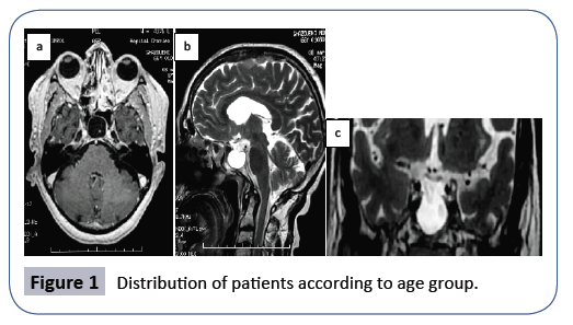
A 67 year old man was referred to our department for headache and left rhinorrhea evolving since 3 months. His family and personal history were unremarkable. Examination showed CSF rhinorrhea. Neurological examination was normal. The ophthalmologic evaluation was normal. Brain MRI showed empty sella (Figure 1). Endocrinological tests were normal. The patient was operated by endoscopic transphenoidal approach and he was symptomatically better.
Discussion
Spontaneous CSF leak is common. In case of empty sella, the elevated intracranial fluid pressure results in bone thinning in sphenoid sinus and cribriform plate [3]. It may eventually cause brain herniation or bone erosion with CSF leakage. Number of hypotheses may explain the cause of primary ESS. Among them, we quote pituitary infarction, pituitary apoplexy and rupture of an intrasellar cyst [4]. A reasonable explanation is that the condition arises in a patient who has an elevation in intracranial pressure associated with an incompetent diaphragma sella which allows the subarachnoid space to be forced into the sella by the hydrostatic pressure and pulsatile movement of CSF. The bony erosion, especially if augmented by increased intracranial pressure, can cause communication of the intrasellar subarachnoid space with the sphenoid sinus. CSF rhinorrhea may be attributed to benign intracranial hypertension, which can be associated with ESS [5]. The site of the leak is usually into the sphenoid sinus but may be through the cribriform plate and can be distinguished after the injection of the intrathecal contrast. Visual changes are noted. They result from herniation of the suprasellar cisterns into the sellar space [4]. This causes downward displacement of the optic nerves, optic chiasm and exposes the optic structures to a more intense CSF pulsation [4].
The MRI represents the gold standard for the diagnosis of the empty sella. It shows a large sella filled with CSF. Because of the high risk of CSF rhinorrhea and infection, the intradural technique was replaced by the extradural technique which represents the current treatment modality [4,6]. Several materials have been suggested for filling the sellar space and reconstruction the sellar floor. As recorded by many authors the fat was preferable to muscle because it results in less necrosis or scar retraction over time [4]. The technique consists of inserting an amount of fat inside the sella which will push the optic structure into their normal position. Extradural transsphenoidal chiasmapexy can be indicated if the optic chiasm herniated inside the sella causing progressive visual abnormalities [7]. They include muscle, fat, dural substitutes, cartilage, bone fragments, ceramic substances, titanium plates...
The endoscopic approach is considered the preferred procedure for treatment of sinus CSF leaks [6,8]. The success rate is estimated between 90 and 95%. It is associated with less complication than the open skull approaches. The surgery consists on separation of the communication from the nose and sinuses to the brain compartment. The endoscope helps to identify the site of CSF leak and to place the grafts precisely [9].
Conclusion
The primary empty sella is generally asymptomatic and incidentally detected, it requires no specific treatment. However, when it is associated with cerebrospinal fluid rhinorhea, surgical treatment will be mandatory. The endoscopic transnasal transphenoidal approach appears to be the best option.
References
- Sage MR, Blumbergs PC (2000) Primary empty sella turcica: a radiological–anatomical correlation. Journal of Medical Imaging and Radiation Oncology 44: 341-348.
- Chi JH, Park SW, Han MS, Lee EK (2011) A Case of Endoscopic Repair of Cerebrospinal Fluid Rhinorrhea Associated with Primary Empty Sella Syndrome. Korean Journal of Otorhinolaryngology-Head and Neck Surgery 54: 727-730.
- Darouassi Y, Mliha TM, Chihani M, Akhaddar A, Ammar H, et al. (2016) Spontaneous cerebrospinal fluid leak of the sphenoid sinus mimicking allergic rhinitis, and managed successfully by a ventriculoperitoneal shunt: a case report. J Med Case Rep 10: 308.
- Fouad W (2011) Review of empty sella syndrome and its surgical management. Alexandria Journal of Medicine 47: 139-147.
- Perez MA, Bialer OY, Bruce BB, Newman NJ, Biousse V (2013) Primary spontaneous cerebrospinal fluid leaks and idiopathic intracranial hypertension. Journal of neuro-ophthalmology: the official journal of the North American Neuro-Ophthalmology Society 33: 330.
- Yadav YR, Parihar V, Janakiram N, Pande S, Bajaj J, et al. (2016) Endoscopic management of cerebrospinal fluid rhinorrhea. Asian J Neurosurg 11: 183.
- Barzaghi LR, Donofrio CA, Panni P, Losa M, Mortini P (2018) Treatment of empty sella associated with visual impairment: a systematic review of chiasmapexy techniques. Pituitary 21: 98-106.
- Presutti L, Mattioli F, Villari D, Marchioni D, Alicandri-Ciufelli M (2009) Transnasal endoscopic treatment of cerebrospinal fluid leak: 17 years’ experience. Acta Otorhinolaryngol Ital 29: 191-196.
- Virk JS, Elmiyeh B, Saleh HA (2013) Endoscopic management of cerebrospinal fluid rhinorrhea: the charing cross experience. J Neurol Surg B Skull Base 74: 61.
Open Access Journals
- Aquaculture & Veterinary Science
- Chemistry & Chemical Sciences
- Clinical Sciences
- Engineering
- General Science
- Genetics & Molecular Biology
- Health Care & Nursing
- Immunology & Microbiology
- Materials Science
- Mathematics & Physics
- Medical Sciences
- Neurology & Psychiatry
- Oncology & Cancer Science
- Pharmaceutical Sciences

