ISSN : 2393-8854
Global Journal of Research and Review
The role of neuroisualization research in neonatal surgery
Abstract
Infant mortality of developmental abnormalities in the world rank third. The severity of the condition of such children at birth is often due to pulmonary-cardiac insufficiency, unstable hemodynamics, which increases the risk of perinatal CNS damage. Recent advances in pediatric neurology, pediatric surgery and neonatology in recent years have identified the possibility of providing emergency care for newborn children with severe developmental anomalies.
Neurosonography is traditionally used as the main method for early diagnosis of structural brain lesions in newborns. The method is widely used in connection with general accessibility, safety and low invasiveness, and is invaluable in diagnosing brain lesions in children in hospital units, transportation of which is impossible for MRT studies due to the severity of the condition.
Information of a higher degree and the nature of the anatomical structures of the brain can be produced by the MRI method, this method is characterized by the absence of radiation load on the body, the possibility of research in various planes without moving the patient, and high tissue contrast. In addition, MRI is a standardized reproducible method, which makes it more objective than other types of neuroimaging, including neurosonography.
Author(s): Igamova S.S
Abstract | PDF
Share This Article
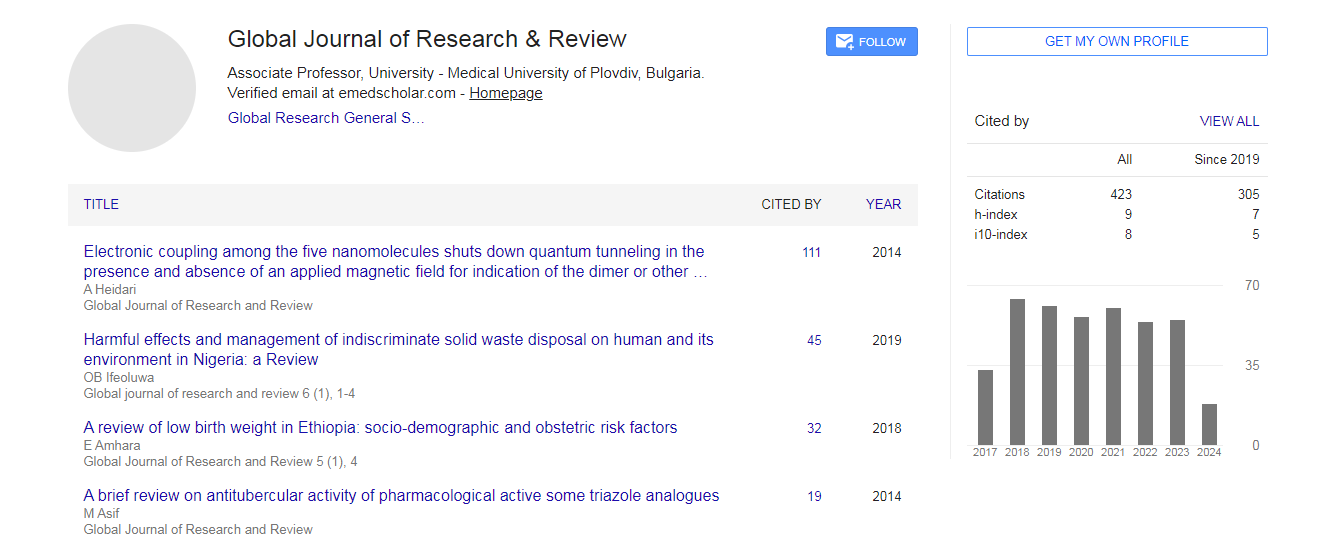
Google Scholar citation report
Citations : 422
Global Journal of Research and Review received 422 citations as per Google Scholar report
Abstracted/Indexed in
- Google Scholar
- Genamics JournalSeek
- China National Knowledge Infrastructure (CNKI)
- Directory of Research Journal Indexing (DRJI)
- Secret Search Engine Labs
Open Access Journals
- Aquaculture & Veterinary Science
- Chemistry & Chemical Sciences
- Clinical Sciences
- Engineering
- General Science
- Genetics & Molecular Biology
- Health Care & Nursing
- Immunology & Microbiology
- Materials Science
- Mathematics & Physics
- Medical Sciences
- Neurology & Psychiatry
- Oncology & Cancer Science
- Pharmaceutical Sciences