Seroprevalence and Spatial Analysis of Canine Visceral Leishmaniasis in the Pantanal Region, Mato Grosso State, Brazil
Álvaro Felipe de Lima Ruy Dias1, Arleana do Bom Parto Ferreira de Almeida1, Felipe Augusto Costantino Seabra da Cruz2, Rafael Rodrigues da Silva2, Juliana Yuki Rodrigues2, Amanda Atsumy Funakawa Otsubo3, Anderson Castro Soares de Oliveira4 and Valéria Régia Franco Sousa1*
1Faculty of Veterinary Medicine, Federal University of Mato Grosso, Cuiabá, Mato Grosso, Brazil
2Veterinary Clinic, Cuiabá, Mato Grosso, Brazil
3National Council for Scientific and Technological Development, Federal University of Mato Grosso, Cuiabá, Mato Grosso, Brazil
4Department of Statistics, Institute of Exact and Earth Sciences, Federal University of Mato Grosso, Cuiabá, Mato Grosso, Brazil
- *Corresponding Author:
- Valéria Régia Franco Sousa
Department of Veterinary Medical Clinic
Faculty of Veterinary Medicine
Federal University of Mato Grosso, Brazil
Tel: 55-65-36158662
Fax: 55-65-36158662
E-mail: valeriaregia27@gmail.com
Received Date: December 18, 2016; Accepted Date: January 03, 2017; Published Date: January 10, 2017
Citation: Dias AFLR, Almeida ABPF, Cruz FACS, et al. Seroprevalence and Spatial Analysis of Canine Visceral Leishmaniasis in the Pantanal Region, Mato Grosso State, Brazil. J Zoonotic Dis Public Health. 2017, 1:3.
Abstract
Introduction: Visceral Leishmaniasis (VL) is a zoonotic disease of public health importance, whose main reservoir is the domestic dog. The purpose of this study was to evaluate the prevalence and spatial distribution of Canine Visceral Leishmaniasis (CVL), as well as factors associated with the disease, in the municipality of Barão de Melgaço.
Methods: Serum samples from 402 dogs in urban and rural areas were subjected to an immunochromatographic rapid test (DPP, TR DPP® LVC–Bio-Manguinhos/FIOCRUZ) and the enzyme-linked immunosorbent assay (ELISA, EIE® LVC–Bio-Manguinhos/FIOCRUZ). An epidemiological questionnaire was used to identify factors associated for CVL using a logistic regression model. The presence of spatial autocorrelation was performed using global and local Moran’s indices.
Results: Seventeen dogs were reactive to both diagnostic tests, indicating a prevalence rate of 4.2% in the municipality, and a statistically significant association with hyperkeratosis and splenomegaly (P<0.05). Through the global Moran index, a density map was built for show the spatial distribution of CVL cases, showing a positive correlation among all the evaluated areas (I<0.25). Local Moran index was used to create a cluster map, identifying areas with high, low and intermediate priority for control of CVL.
Conclusions: The prevalence of CVL in Barão de Melgaço was of 4.2%, being characterized as area with moderate transmission and receptive to the occurrence of human cases. In this study, the use of spatial analysis stood out as a strategic tool in the control of CVL, enabling different action strategies to be applied in each region.
Keywords
Dog; Epidemiology; Georeferencing; Leishmania chagasi; Zoonotic disease
Introduction
Brazil is among the six countries most affected by Visceral Leishmaniasis (VL) [1]. It is estimated that about 90% of VL cases in the New World occur in Brazil, where it is rapidly spreading to the country’s five regions [2,3]. In the New World, this zoonotic disease is caused by the protozoan Leishmania chagasi (syn. Leishmania infantum) transmitted to vertebrate hosts through the bite of infected dipterans [1,4].
Dogs, which are considered the main hosts and reservoirs of the disease in urban environments, are highly susceptible to infection, characterized by intense cutaneous parasitism, which favors infection of the vector and, hence, transmission to humans [5,6].
The proximity of Barão de Melgaço to regions endemic for CVL, such as Cuiabá [7], Rondonópolis [8], Poxoréo [9], Jaciara and Várzea Grande [10], allied to the region’s climatic and physiographic characteristics, which favour the presence of the vectors Lutzomyia longipalpis and L. cruzi already reported in the municipality [11], can contribute to the persistence and dissemination of this disease. This means the region represents a risk to the local population and to tourists from different parts of the world, which come in search of the natural attractions of North Pantanal fauna and flora.
Given the importance of dogs in the epidemiological cycle of VL and the tourist potential of Barão de Melgaço, this study aimed to evaluate the seroprevalence and spatial distribution of CVL in Barão de Melgaço, associating the disease to environmental and individual factors.
Methods
Study design
A descriptive cross-sectional study was conducted in the city of Barão de Melgaço, located in the Pantanal region of the state of Mato Grosso, Brazil (coordinates: latitude 16°11'40''S and 55°58'03''W). The minimum sample size was estimated at 283 dogs, considering a dog to human ratio of 7:1, at the estimated population of 7,526 in 2015 [12], using a confidence level of 95%, acceptable error of 5% and prevalence of 50% [3]. Samples were collected from dogs in all the urban neighbourhoods, and from dogs in 11 rural communities.
Samples and serological testing
Blood samples were drawn from dogs of both sexes, different breeds, and ages equal to or older than six months, which had been previously examined and classified as symptomatic or asymptomatic [13]. At each residence, the dog’s owner was asked to fill out an epidemiological questionnaire containing information about the dog’s breed, sex, age, fur length, function, the place it occupies in the house, its access to the street, proximity of the house to forest and rivers, location of the house, the presence of other animals and/or chicken coops or pig sties, vegetation around the house, and garbage collection.
The dogs were serologically diagnosed using the tests recommended by the Ministry of Health (MS): DPP for screening and ELISA for confirmation. Both tests were performed according to the instructions of the manufacturer, Bio-Manguinhos/FIOCRUZ.
The DPP kit for rapid detection of K26/K39-specific antibodies was performed as a screening tool at the Laboratory of Leishmaniasis, Department of Clinic Veterinary Medicine, The Federal University of Mato Grosso (CLIMEV-UFMT). The sample was considered positive if two red lines appeared and negative if only one red line appeared. Subsequently, the samples positive on DPP were subjected to ELISA, which employs a soluble antigen from promastigote forms of L. major-like parasites. This serology was performed in the Laboratory of Leishmaniasis of the CLIMEV-UFMT. The sample was considered positive when the ELISA reading was higher than the cut-off point. The results were obtained by a combination of the two tests.
All of the laboratorial procedures were executed according to the Principles of Good Laboratory Practices [3] for the protection of the manipulator, sample and environment.
Data analysis
Two-step logistic regression, i.e., univariate and multivariate analysis, was employed to correlate the seroprevalence of anti-L. chagasi antibodies in dogs and the independent variables. Variables presenting a chi-square P ≤ 0.20 in the univariate analysis were selected for the multivariate analysis, and P ≤ 0.05 were considered significant. The correlation between the two diagnostic tests was established based on the Kappa test [14]. The tests were performed in R 3.3.0 using the R Commander package [15].
Spatial analysis
In the spatial analysis, the global Moran index was employed as a measure of spatial autocorrelation in the various census tracts of Barão de Melgaço, this index provides a single value, potentially varying from -1 to 1. As for the local Moran index, the tracts were classified into four quadrants based on the Moran scatter plot, where: quadrant HH (+/+) and quadrant LL (-/-) are areas with a positive spatial association, while quadrant HL (+/-) and quadrant LH (-/+) are areas with a negative spatial association. Three areas were defined with different CVL control priorities: High priority– quadrant HH (+/+), low priority–quadrant LL (-/-), and intermediate priority–quadrants HL (+/-) and LH (-/+). A representative map was used to examine the areas with statistically significant spatial autocorrelations (P ≤ 0.01) [16]. The analyses were performed in R 3.3.0 using the R Commander package [15].
Ethics
This study was conducted in accordance with the ethical principles approved by the Ethics Committee on Animal Use (CEUA)–UFMT under protocol number 23108.014950/11-5. The dog owners were informed of the research objectives and were required to sign an informed consent form before sample and data collection.
Results
Of the 402 evaluated dogs (248 from urban and 154 from rural areas), assuming parallel results, 94 samples tested positive in at least one of the serological assays, with a seroprevalence of 23.4% (95% CI: 19.39 to 27.89). In an individual analysis, the DPP screening test showed a seroprevalence of 19.4% (95% CI: 15.72 to 23.68, 78/402), while the confirmatory test, ELISA, showed a seroprevalence of 8.2% (95% CI: 5.8 to 11.45, 33/402).
Based on the combined results and serial protocol stipulated by the MS, 17 samples were positive in both tests, with a seroprevalence of 4.2% (95% CI: 2.56 to 6.82). Rural dogs (4.5%) showed a 0.5% higher seroprevalence than urban dogs (P=0.803). The Kappa test of the DPP and ELISA results showed a slight agreement of 21% (95% CI: 0.12 to 0.30).
In general, this study contained predominantly male dogs (57%, 229/402), mixed breed dogs (90%, 362/402), adults (92.8%, 371/402), unneutered (96%, 373/402), and short-haired dogs (89.8%, 361/402), and no statistical association was found between their characteristics and the prevalence of infection (P>0.05) (Table 1). In the clinical examination, 14 (82.35%) seropositive dogs showed clinical signs suggestive of CVL (P=0.064) (Table 1), the most common ones being lymphadenopathy (47.1%) and skin lesions (41.2%), followed by splenomegaly and onychogryphosis (35.3%), ophthalmopathy and alopecia (23.5%). Scaling and hyperkeratosis (11.8%), ear tip ulcers and periocular alopecia (5.9%) were less frequent.
| Variables | DPP and ELISA | P-value | OR (95% CI)* | |
|---|---|---|---|---|
| Positive | Negative | |||
| n (%) | n (%) | |||
| Breeda | ||||
| CRD | 1 (2.5) | 39 (97.5) | 1 | ** |
| SRD | 16 (4.4) | 346 (95.6) | ||
| Sex | ||||
| Male | 11 (4.8) | 218 (95.2) | 0.51 | 1.4 (0.46-4.72) |
| Female | 6 (3.5) | 167 (96.5) | 1 | |
| Age groups | ||||
| Indefinite age | 4 (6.25) | 60 (93.75) | 0.671 | ** |
| ≤1 | 1 (3.2) | 30 (96.8) | ||
| ≥1-3 | 4 (3.6) | 106 (96.4) | ||
| ≥3-5 | 3 (2.7) | 110 (97.3) | ||
| ≥5 | 5 (6.0) | 79 (94.0) | ||
| Fur length | 1 | ** | ||
| Short | 16 (4.4) | 345 (95.6) | ||
| Long | 1 (2.4) | 40 (97.6) | ||
| Clinical status | ||||
| Asymptomatics | 3 (1.9) | 154 (98.1) | 0.064 | 1 |
| Symptomatic | 14 (5.7) | 231 (94.3) | 3.1 (0.84-17.12) | |
| House role | ||||
| Guard | 6 (3.9) | 148 (96.1) | 0.95 | ** |
| Company | 8 (4.4) | 172 (95.6) | ||
| Both | 2 (4.1) | 47 (95.9) | ||
| Hunt | 1 (5.3) | 18 (94.7) | ||
| Dog habitat | ||||
| Intradomestic | - | 10 (100.0) | 0.609 | ** |
| Peridomiciliar | 17 (4.8) | 336 (95.2) | ||
| Both | - | 39 (100.0) | ||
| Proximity to: | ||||
| Forest | 6 (4.4) | 131 (95.6) | 0.952 | ** |
| Wasteland | 1 (92.6) | 37 (97.4) | ||
| River | 3 (3.3) | 89 (96.7) | ||
| Any combination | 5 (5.2) | 91 (94.8) | ||
| Neither | 2 (5.1) | 37 (94.9) | ||
| Residence type | ||||
| Urban | 10 (4.0) | 238 (96.0) | 0.803 | 1 |
| Rural | 7 (4.5) | 147 (95.5) | 1.13 (0.35-3.38) | |
*Odds Ratio; 95% CI: 95% Confidence interval **Has not been determined due to lack of categories, null or very low frequencies aCRD–With defined race; SRD–Without defined race
Table 1: Univariate analyses for variables considered for the study of risk factors associated with canine visceral leishmaniasis in 402 dogs from Barão de Melgaço, State of Mato Grosso, Brazil.
In the univariate analysis for anti-L. chagasi antibodies, the variables of splenomegaly, ophthalmopathy, periocular alopecia and hyperkeratosis showed values of P<0.20 and were selected for multivariate analysis. This analysis indicated that the main factors associated with canine infection were the presence of hyperkeratosis and splenomegaly, which were, respectively, 16.19 and 4.05 times more likely to be present in dogs seropositive for CVL than in those without these clinical signs (Table 2).
| Variables | Dogs | Univariate analysis | Multivariate analysis | |||
|---|---|---|---|---|---|---|
| n | Positive (%) | X2 | P | P | OR (95% CI)* | |
| Splenomegaly | ||||||
| Yes | 63 | 6 (9,5) | 5,29 | 0,017 | 0,014 | 4,05 (1,32-12,43) |
| No | 355 | 11 (3,1) | ||||
| Lymphadenopathy | ||||||
| Yes | 164 | 8 (4,9) | 0,288 | 0,591 | - | - |
| No | 238 | 9 (3,8) | ||||
| Onychogryphosis | ||||||
| Yes | 32 | 3 (9,4) | 2,273 | 0,131 | 0,997 | 1,0 (0,19-5,09) |
| No | 170 | 14 (3,8) | ||||
| Ocular disturbances | ||||||
| Yes | 37 | 4 (10,8) | 4,359 | 0,036 | 0,486 | 1,63 (0,41-6,50) |
| No | 365 | 13 (3,6) | ||||
| Periocular alopecia Yes | ||||||
| No | 1 | 1 (100,0) | 22,70 | < 0,01 | 0,991 | ** |
| 401 | 16 (4,0) | |||||
| Hyperkeratosis | ||||||
| Yes | 4 | 2 (50,0) | 20,89 | <0,01 | 0,033 | 16,19 (1,24-210,6) |
| No | 398 | 15 (3,8) | ||||
| Domestic animals | ||||||
| Yes | 395 | 11 (3,7) | 0,684 | 0,408 | - | - |
| No | 107 | 6 (5,6) | ||||
| Animal husbandry | ||||||
| Yes | 227 | 10 (4,0) | 0,04 | 0,841 | - | - |
| No | 175 | 7 (4,0) | ||||
| Access to the street | ||||||
| Yes | 297 | 12 (4,0) | 0,099 | 0,752 | - | - |
| No | 105 | 5 (4,8) | ||||
| Vegetation | ||||||
| Yes | 363 | 15 (4,1) | 0,086 | 0,769 | - | - |
| No | 39 | 2 (5,1) | ||||
| Garbage collection | ||||||
| Yes | 244 | 10 (4,1) | 0,026 | 0,871 | - | - |
| No | 158 | 7 (4,4) | ||||
* Odds ratio; 95% CI, 95% Confidence interval. ** Has not been determined due to lack of categories, null or very low frequencies
Table 2: Univariate and multivariate analyses to detect risk factors associated with the DPP and ELISA positivity of canine visceral leishmaniasis in dogs from Barão de Melgaço, State of Mato Grosso, Brazil.
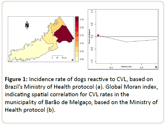
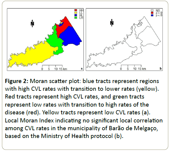
A choropleth map (Figure 1a) was created to ascertain the incidence rates of CVL in the census tracts of Barão de Melgaço. On this map, dark areas indicated a higher concentration of cases appearing in the north-south direction of the municipality, while light areas in the central and western part indicated the absence or a lower concentration of cases. To identify the spatial correlation structure that best describes the data, a Moran correlogram was built using the global Moran index (Figure 1b), which revealed a positive correlation for all the evaluated tracts, since the value was above zero, although the correlation was small or zero (I<0.25). A cluster map based on the Moran scatter plot (Figure 2a) showed that only one tract presented high priority–HH (+/+) and three presented low priority–LL (-/-), indicating points of positive spatial association, i.e., tracts with neighbors showing similar values. As for the other tracts, four showed intermediate priority–HL (+/-), LH (-/+), indicating points of negative spatial association, i.e., contiguous census tracts with different values. The local Moran index (Figure 2b) indicated that there was no significant local correlation.
Figure 2: Moran scatter plot: blue tracts represent regions with high CVL rates with transition to lower rates (yellow). Red tracts represent high CVL rates, and green tracts represent low rates with transition to high rates of the disease (red). Yellow tracts represent low CVL rates (a). Local Moran Index indicating no significant local correlation among CVL rates in the municipality of Barão de Melgaço, based on the Ministry of Health protocol (b).
Discussion
This epidemiological survey of canine leishmaniasis, which revealed a 4.2% prevalence of anti-L. chagasi antibodies, was motivated by the proximity of Barão de Melgaço to cities endemic for CVL. Similar results were found in studies conducted in different regions of Brazil. CVL prevalence rates of 3.4% were reported in the municipality of Cuiabá-MT [17], of 4.63% in Divinópolis-MG [18], and of 4.5% in Presidente Prudente-SP [19]. Despite these similar results, the literature describes prevalence rates ranging from 3.4-67% [17,20] in different regions of the country.
Serological tests are important tools in the diagnosis of CVL, and are often used in epidemiological surveys on canines.
Therefore, agreement between the two tests recommended by the MS is very important, in order to ensure that animals testing false-negative in the DPP be subjected to ELISA, which was not observed in this study and was also described by Fonseca et al. [21] and in the meta-analysis of Peixoto et al. [22]. According to the literature, low concordance between tests may be caused by factors such as areas where transmission is still incipient, CVL prevalence rates, and the fact that concordance between diagnostic tests tend to be low [22]. Moreover, dogs infected with L. chagasi present different humoral immune responses to various antibodies at different stages of the disease [23,24]. This means that serological tests may present different degrees of sensitivity, depending on the clinical course of the infection, genetic variability of the host, and specific immune responses [25,26].
Other factors also interfere in the serological diagnosis, such as the type of antigen used for the diagnosis of antibodies [23,27]. In this context, the DPP assay uses recombinant proteins, and despite the technique’s high sensitivity (98%) and specificity (96%) in symptomatic animals, it shows low sensitivity (47%) in asymptomatic dogs [25]. On the other hand, the ELISA, which uses L. major-like crude antigens, provides varying rates of sensitivity (8-100%) and specificity (60-100%) in the diagnosis of CVL due to antigenic similarities between Leishmania species [22,28]. Both subject to cross-reactions and false positive results in overlapping areas of trypanosomatids [28,29].
In this study, symptomatic dogs accounted for 82.35% of the seropositive dogs. This high percentage corroborates results reported in the literature that associate the positivity of serological tests with the severity of the disease [25]. Nevertheless, among animals infected with L. chagasi, asymptomatic animals are usually more prevalent in epidemiological studies [30]. Therefore, further research should be encouraged to devise techniques that are more sensitive to asymptomatic animals. The clinical signs observed in this study were the same as those described in other epidemiological studies of CVL [17], and the univariate and multivariate analysis revealed a positive correlation between hyperkeratosis and splenomegaly and the presence of the disease. This is an important finding in the clinical analysis of animals suspected of having the disease, in view of the large number of its clinical symptoms.
Spatial analysis tools have been increasingly used in studies of VL, and have been the focus of research in different regions of Brazil [18,31]. Thus, epidemiological studies that include spatial analysis can be used in VL surveillance and control programs to identify epidemiologically important areas and to prioritize municipalities that require the adoption of control measures [17,31].
In this context, the incidence of CVL was distributed unevenly in the various tracts of the municipality, with some tracts showing a high concentration of cases and others showing no CVL or a lower concentration of the disease. Only one census tract was identified as high priority for control of the disease. This variation in prevalence rates in the municipality may be ascribed to the different characteristics of each area in Barão de Melgaço, such as the population’s socioeconomic status, agglomerations of animals and humans, poor basic sanitation, and houses usually located near rivers (humid environments) and forests. It should be noted that the only census tract considered as high priority contains both urban and rural areas, and because the municipality is located in the Pantanal wetlands, it is subjected to the annual flood cycle, which means that 98% of its territory is flooded during the rainy season from January to March. As a result, the population tends to migrate from the rural to the urban area, thus favoring the spread of the disease to the urban environment, since sick animals coming from rural areas can contribute to urbanize the disease. Moreover, the decreasing areas of dry land may lead to greater contact between wildlife reservoirs of the disease and the population, and the accumulation of organic matter favours increased vector density. Thus, the municipality may be in the process of urbanization of the disease, because unlike other Brazilian municipalities with larger urban populations, a large part of the population of Barão de Melgaço is rural, which justifies this hypothesis.
Conclusion
The results of this study revealed a CVL prevalence rate of 4.2% in urban and rural areas of the municipality of Barão de Melgaço, being characterized as area with moderate transmission and receptive to the occurrence of human cases. Hyperkeratosis and splenomegaly were identified as factors associated with CVL. The spatial analysis methodology allowed determining priority control areas for CVL, which should be the focus of prevention and control measures.
Acknowledgements
The authors would like to thank the Secretary of State for Health of the State of Mato Grosso, Municipal Secretary of Health of Barão de Melgaço, and Fiocruz (Bio-Manguinhos), for providing the kits used in this research. This work was supported, in part, by grants from the Coordination of Improvement of Higher Education Personnel, Foundation for Research Support of the State of Mato Grosso (154220/2014-3), and National Council for Scientific and Technological Development (445998/2014-8).
References
- https://www.who.int/mediacentre/factsheets/fs375/en/
- Alvar J, Vélez ID, Bern C, Herrero M, Desjeux P, et al. (2012) Leishmaniasis worldwide and global estimates of its incidence. PLoS ONE 7: e35671.
- MS (2014) Ministry of Health: Secretariat of Health Surveillance, Department of Epidemiological Surveillance. Manual of Surveillance and Control of Visceral Leishmaniasis. Publisher of the Ministry of Health Brazil, Braasília-DF.
- Romero GA, Boelaert M (2010) Control of visceral leishmaniasis in Latin America - a systematic review. PLoS Neglect Trop D 4: e584.
- Menezes-Souza D, Corrêa-Oliveira R., Guerra-Sá R, Giunchetti RC, Teixeira-Carvahlo A, et al. (2011) Cytokine and transcription fator profiles in the skin of dogs narueally infected by Leishmania (Leishmania) chagasi presenting distinct cutaneous parasite density and clinical status. Vet Parasitol 177: 39-49.
- Belo VS, Werneck GL, Barbosa DS, Simões TC, Nascimento BW, da Silva ES, Struchiner CJ (2013) Factors associated with visceral leishmaniasis in the américas: a systematic review and meta-analysis. PLos Neglect Trop D 7: e2182.
- Almeida ABPF, Sousa VRF, Cruz FACS, Dahroug MAA, Figueiredo FB, et al. (2012) Canine visceral leishmaniasis: seroprevalence and risk factors in Cuiabá, Mato Grosso, Brazil. Rev Bras Parasitol V 21: 359-365.
- Duarte JLS (2010) Epidemiological aspects of visceral leishmaniasis in the Municipality of Rondonópolis, Mato Grosso, 2003 - 2008.
- Azevedo MAA, Dias AKK, Paula HB, Perri SHV, Nunes CM (2008) Evaluation of canine visceral leishmaniasis in Poxoréo, Mato Grosso State, Brazil. Rev Bras Parasitol V 17:123-127.
- Mestre GLC, Fontes CJFA (2007) The expansion of the visceral leishmaniasis epidemic in the State of Mato Grosso, 1998-2005. Rev Soc Bras Med Trop 40: 42-48.
- Missawa NA, Veloso MAE, Maciel GBML, Michalsky ÉM, Dias ES (2011). Evidence of visceral leishmaniasis transmission by Lutzomyia cruzi in the municipality of Jaciara, State of Mato Grosso, Brazil. Rev Soc Bras Med Trop 44: 76-78.
- https://cod.ibge.gov.br/4T6
- Solano-Gallego L, Koutinas A, Miró G, Cardoso L, Pennisi MG, et al. (2009) Directions for the diagnosis, clinical staging, treatment and prevention of canine leishmaniosis, Vet Parasitol 165: 1-18.
- Landis JR, Koch GG (1977) The measurement of observer agreement for categorical data. Biometrics 33: 159-174.
- Fox J (2005) The R Commander: A basic statistics graphical user interface to R. Journal of Statistical Software 14: 1-42.
- Atanaka-Santos M, Souza-Santos R, Czeresnia D (2007) Spatial analysis for stratification of priority malária control areas, Mato Grosso State, Brazil. Cad Saude Publica 23: 1099-1112.
- Almeida ABPF, Faria RP, Pimentel MFA, Dahroug MAA, Turbino NCMR, et al. (2009) Seroepidemiological survey of canine leishmaniasis in endemic areas of Cuiabá, Mato Grosso State. Rev Soc Bras Med Tro 42: 156-159.
- Teixeira-Neto RG, da Silva ES, Nascimento RA, Belo VS, Oliveira CL, et al. (2014) Canine visceral leishmaniasis in an urban setting of Southeastern Brazil: an ecological study involving spatial analysis. Parasite Vector 7:485.
- D'Andrade LA, Fonseca ES, Prestes-Carneiro LE, Guimarães RB, Yamashita RC, et al. (2015) The shadows of a ghost: a survey of canine leishmaniasis in Presidente Prudente and its spatial dispersion in the western region of São Paulo state, an emerging focus of visceral leishmaniasis in Brazil. BMC Vet Res 11: 273.
- Barbosa DS, Rocha AL, Santana AA, Souza CSF, Dias RA, et al. (2010) Seroprevalence and epidemiological variables associated with canine visceral leishmaniasis in an endemic area in the city of São Luís, Maranhão, Brazil. Brazilian Animal Science 11: 653-659.
- Fonseca AM, Faria AR, Rodrigues FTG, Nagem RAP, Magalhães RDM, et al. (2014) Evaluation of three recombinant Leishmania infantum antigens in human and canine visceral leishmaniasis diagnosis. Acta Trop 137: 25-30.
- Peixoto HM, de Oliveira MRF, Romero GAS (2015) Serological diagnosis of canine visceral leishmaniasis in Brazil: systematic review and meta-analysis. Tropical Medicine and International Health 20:334-352.
- Porrozzi R, Santos Da Costa MV, Teva A, Falqueto A, Ferreira AL, et al. (2007) Comparative evaluation of enzyme-linked immunosorbent assays based on crude and recombinant leishmanial antigens for serodiagnosis of symptomatic and asymptomatic Leishmania infantum visceral infections in dogs. Clin Vaccine Immunol 14: 544-548.
- Falqueto A, Ferreira AL, Dos Santos CB, Porrozzi R, da Costa MVS, et al. (2009) Cross-sectional and longitudinal Epidemiologic Survevy of Human and Canine Leishmania infantum Visceral Infections in an Endemic Rural Area of Southeast Brazil (Pancas, Espírito Santo). The American Society of Tropical Medicine and Hygiene 80: 559-565.
- Grimaldi GJr, Teva A, Ferreira AL, dos Santos CB, Pinto LD, et al. (2012) Evaluation of a novel chromatographic immunoassay based on Dual-Path Platform technology (DPP® CVL rapid test) for the serodiagnosis of canine visceral leishmaniasis. T Roy Soc Trop Med H 106: 54-59.
- Quinnell RJ, Carson C, Reithinger R, Garcez LM, Courtenay O (2013) Evaluation of rK39 rapid diagnostic tests for canine visceral leishmaniasis: longitudinal study and meta-analysis. PLoS ONE 7: e1992.
- Fraga DBM, da Silva ED, Pacheco LV, Borja LS, de Oliveira IQ, et al. (2014) A multicentric evaluation of the recombinant Leishmania intantum antigen-based immunochromatographic assay for the serodiagnosis of canine visceral leishmaniasis. Parasite Vector 7: 136.
- Oliveira E, Saliba JW, Oliveira D, Dias ES, Paz GF (2016) A prototype of the direct agglutination test kit (DAT-Canis) for the serological diagnosis of canine visceral leishmaniasis. Vet Parasitology 221: 9-13.
- Alves AS, Couta-Confort EM, Figueiredo FB, Oliveira RVC, Schubach AO, et al. (2012) Evaluation of serological cross-reactivity between canine visceral leishmaniasis and natural infection by Trypanosoma caninum. Res Vet Sci 93: 1329-1333.
- Queiroz PV, Monteiro GR, Macedo VP, Roch MA, Batista LM, et al. (2009) Canine visceral leishmaniasis in urban and rural areas of Northeast Brazil. Res Vet Sci 86: 267-273.
- Barbosa DS, Belo VS, Rangel MES, Werneck GL (2014) Spatial analysis for identification of priority areas for surveillance and control in a visceral leishmaniasis endemic area in Brazil. Acta Trop 131: 56-62.
Open Access Journals
- Aquaculture & Veterinary Science
- Chemistry & Chemical Sciences
- Clinical Sciences
- Engineering
- General Science
- Genetics & Molecular Biology
- Health Care & Nursing
- Immunology & Microbiology
- Materials Science
- Mathematics & Physics
- Medical Sciences
- Neurology & Psychiatry
- Oncology & Cancer Science
- Pharmaceutical Sciences


