Primary Ovarian Hydatid Cyst: A Case Report and Review of Literature
Fatnassi R*, Turki E, Majdoub W, Hammami S, Hajji M
Department of Obstetrics and Gynecology, University Hospital Ibn, Eljazzar of Kairouan, Tunisia
- *Corresponding Author:
- Fatnassi R
Department of Obstetrics and Gynecology University Hospital
Ibn El Jazzar 3140 Kairouan, Tunisia
Tel: 34620866274
E-mail: ridha.fatnassimohamed@rns.tn
Received date: June 07, 2017; Accepted date: June 22, 2017; Published date: June 28, 2017
Citation: Fatnassi R, Turki E, Majdoub W, Hammami S, Hajji M (2017) Primary Ovarian Hydatid Cyst: A Case Report and Review of Literature. Insights Reprod Med. Vol.1 No.1:5
Copyright: © 2017 Fatnassi R, et al. This is an open-access article distributed under the terms of the Creative Commons Attribution License, which permits unrestricted use, distribution, and reproduction in any medium, provided the original author and source are credited.
Abstract
Hydatidosis is an endemic parasitosis in Tunisia. Primary ovarian localization is exceptional and doubtful. The treatment of choice is surgery consisting, when possible, in a radical laparotomic cystectomy. We report a case of a 32 year old woman affected by right ovarian hydatid cyst that has been treated by surgical cystectomy with good outcome. Etiologic factors, pathophysiology, clinical presentation, diagnosis and main treatment are discussed referring to literature.
Keywords
Hydatidosis; Cyst; Ovarian; Pelvic; Laparotomy
Introduction
Hydatid disease is an endemic zoonosis in Tunisia seen in 15 per 100000 surgical cases [1]. It is caused by the development of a larval stage of Echinococcus granulosus. The parasite embryo is developing in intestinal tract of dogs usually considered as definitive hosts. Humans are an intermediate host who may become infected after ingestion of material contaminated with Echinococcus eggs. After oral contamination, eggs get into blood circulation from the human intestine and may achieve any organ [1-3]. Whereas liver and lung are the most commonly affected accounting for up to 80% all hydatid cyst, the disease may involve every organ. Pelvis is considered as an extremely rare condition occurring in 0.3% to 4.27% of cases. Primitive pelvic hydatid cyst remains a very rare site of cyst formation and deceptive leading to a difficult pre-operative diagnosis [3]. Ovary seems to be the most frequently genital organ involved and constitutes 0.2% of different hydatid disease locations [4-6].
Case Report
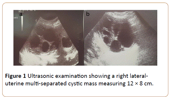
A 32 year old woman gravida living in rural area and without previous medical history presented for our department complaining of isolate chronic pelvic pain evolving since 10 months. She mentioned that she was living in rural areas and was in close contact with dogs and sheep. A physical examination did not reveal any abnormalities. A pelvic examination showed a palpably mass in right midline area of approximately 10 cm in diameter, firm, mobile and painful. Pelvic ultrasonography was ordered and it revealed a right lateral-uterine multi-separated cystic mass measuring 12 × 8 cm that was thought to be most likely from the right ovary without pelvic free fluid (Figures 1a and 1b).
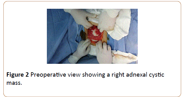
The other abdominal organs were normal. The diagnosis of ovarian mucoid cyst was suggested. White blood cell count, haemoglobin level and haematocrit were normal. Biochemical serum parameters showed revealed no eosinophila. Tumoral CA 19-9 and CA 125 were negatives. Hydatid serology test was performed on the patient’s serum and was positive. Based on these results with the diagnosis of a right ovarian hydatid cyst, an emergency laparotomy was scheduled. Midline incision was performed and showed a right adnexal cystic mass (Figure 2).
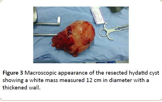
The mass was adherent to the caecum, peritoneum and omentum. Macroscopic examination showed a white mass measuring 12 cm in diameter with a thickened wall as demonstrated in Figure 3.
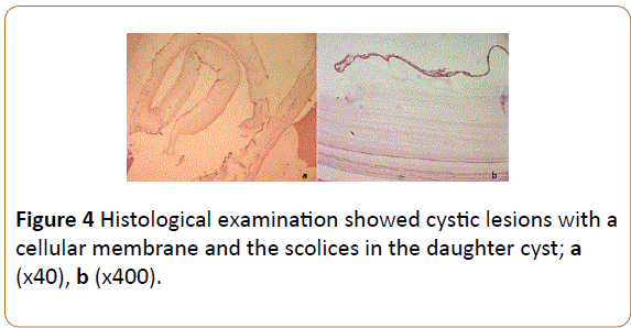
No other intra-abdominal and pelvic cysts were found and uterus and left ovary were normal. A complete cyst wall dissection was performed and peritoneal toilet with hypertonic saline of the cavity was done. The mass was sent for histopathology which revealed multiple small thin-walled daughter cysts. Microscopic diagnosis showed cystic lesions with a cellular membrane and the scolices in the daughter cyst as demonstrated in Figures 4a and 4b.
No others cyst were revealed in chest radiography and abdominal ultrasonography concluding to a primitive characteristic of the ovarian hydatid cyst. The patient did well postoperatively and she was discharged on the 6th postoperative day to be followed up in the outpatient clinic without albendazole prescription. Chest radiograph and abdominal ultrasonography performed 3 and 6 months post operatively were normal. The patient was asymptomatic one year later and hydatid serology test was negative concluding to no recurrence.
Discussion
Hydatid cyst is a parasitic infection which spreads to humans by contagion as en result of close contact with dogs and sheeps [1]. In agreement with previous reports, our patient was from rural areas and was in close contact with those animals.
Primary hydatid cyst is considered when no other sites of occurrence are detected and it is commonly involving liver and lung. Although the mechanism of primary pelvis involvement has not been clearly explained, it has been suggested that scolex gains access to the pelvis by the lymphatic or haematogenous way [7]. We report a case of primary and solitary ovarian hydatid cyst, as no other cyst in the body was detected.
Pelvic hydatid cyst symptomatology is mostly nonspecific and doubtful. Although symptoms are frequently absent, pelvic pain and abdominal mass are the most complained ones. The other reported symptoms include compression of surrounding organs, infertility and menstruation irregularities [8]. In our case, patient is complaining of isolate chronic pelvic pain.
Imaging tools are important to pelvic hydatid disease diagnosis because of the lack of specific clinical signs. Pelvic ultrasound has a low cost and a high sensitivity and constitutes the method of choice. It allows establishing five sonographic types of hepatic hydatid disease which are as follows [9]:
Type I
Purely and unilocular cyst.
Type II
Cyst with a floating membrane.
Type III
Fluid collection with daughter vesicles.
Type IV
Heterogeneous mass.
Type V
Calcified cyst.
Pelvic and abdominal computed tomography has high cost. Nevertheless, it allows an interest evaluation of the features and extension of cystic masses. Because of its high cost, Pelvic magnetic resonance imaging (MRI) is reserved for few cases. Thanks to its performance, MRI may be useful to classify cysts with high specificity and to recognize differential diagnosis commonly including myxoid tumor such as myxoid neurofibroma and angiomyxoma. Chest radiographs are useful to detect associated pulmonary hydatid cyst [10]. In our case, pelvic hydatid cyst diagnosis was suggested thanks to ultrasound. Serologic tests are lowly sensitive and specific to detect Echinococcus antibody in case of pelvic hydatid disease with a risk of false negatives cases. Eosinophila is not reliable and is detected in 33 to 53% of cases [6]. In our case, hydatid serology test was performed and was positive. Nevertheless, blood parameters showed no eosinophila.
The optimal treatment of pelvic hydatid cyst remains surgery. The main principle of surgical treatment is to practise a total resection which allows avoiding perioperative rupture of the cyst [6,11]. In our case, successful complete cystectomy was performed without complications.
The recurrence ratio after surgical treatment has been reported to be about 2% [12]. Albendazole has been considered as the main medical treatment which may reduce recurrence. Nevertheless, most authors suggest that postoperative medical treatment is useless in cases of isolated hydatid cyst without preoperative complications [13]. In our case, albendazole has been no used since ovarian hydatid cyst was isolated and total resection was performed without complications.
Conclusion
Hydatid disease is still considered as a public health problem in Tunisia especially in rural areas. Primary ovarian hydatid cyst remains an exceptional disease often of fortuitous discovery. Diagnosis is frequently difficult because of non-specific clinical presentation. Pelvic ultrasound constitutes the method of choice to elucidate diagnosis. The treatment of choice is surgical consisting preferentially in total resection.
References
- Bellil S, Limaiem F, Bellil K, Chelly I, Mekni A, et al. (2009) Epidemiology of extra pulmonary hydatid cysts: 265 Tunisian cases. Med Mal Infect 39: 341-343.
- Kiresi DA, Karabacakog˘lu A, Odev K, Karaköse S (2003) Uncommon locations of hydatid cysts. Acta Radiol 44: 622-636.
- Hangval H, Habibi H, Moshref A, Rahimi (1979) A: Case report of an ovarian hydatid cyst. J Trop Med Hyg 82: 34.
- Aksu MF, Budak E, Ince U, Aksu C (1997) Hydatid cyst of the ovary. Arch Gynecol Obstet 261: 51-53.
- Diaz-Recasens J, Garcia-Enguidanos A, Munoz I, Sainz de la Cuesta R (1998) Ultrasonographic appearance of an Echinococcus ovarian cyst. Obstet Gynecol 91: 841-842.
- Baba A, Chaieb A, Khairi H, Keskes J (1991) Epidemiological profile of pelvic hydatidosis: 15 cases [in French]. J Gynecol Obstet Biol Reprod (Paris) 20: 657-660.
- Uchikova E, Pehlivanov B, Uchikov A, Shipkov C, Poriazova E (2009) A primary ovarian hydatid cyst. Aust N Z J Obstet Gynaecol 49: 441-442.
- Dede S, Dede H, Caliskan E, Demir B (2002) Recurrent pelvic hydatid cyst obstructing labor, with a concomitant hepatic primary. A case report. J Reprod Med 47: 164-166.
- Gharbi HA, Hassine W, Brauner MW, Dupuch K (1981) Ultrasound examination of hydatid liver. Radiology 139: 459-463.
- Varedi P, Saadat Mostafavi SR, Salouti R, Saedi D, Nabavizadeh SA, et al. (2008) Hydatidosis of the pelvic cavity: A big masquerade. Infect Dis Obstet Gynecol 2008: 782621.
- Gamoudi A, Ben Romdhane K, Farhat K, Khattech R, Hechiche M, et al. (1995) Ovarian hydatic cyst: 7 cases [in French]. J Gynecol Obstet Biol Reprod (Paris) 24: 144-148.
- Bull World Health Organ (1996) Guidelines for treatment of cystic and alveolar echinococcosis in humans. WHO informal working group on Echinococcosis. 74: 231-242.
- Akbulut S, Senol A, Ekin A, Bakir S, Bayan K, et al. (2010) Primary retroperitoneal hydatid cyst: report of 2 cases and review of 41 published cases. Int Surg 95: 189-196.
Open Access Journals
- Aquaculture & Veterinary Science
- Chemistry & Chemical Sciences
- Clinical Sciences
- Engineering
- General Science
- Genetics & Molecular Biology
- Health Care & Nursing
- Immunology & Microbiology
- Materials Science
- Mathematics & Physics
- Medical Sciences
- Neurology & Psychiatry
- Oncology & Cancer Science
- Pharmaceutical Sciences
