Hearing Organ Assessment using Pure-Tone Audiometry in Children and Adolescents with Peripheral Paralysis of the Facial Nerve
Wacław Kopala1* and Andrzej Kukwa2
1The Provincial Children's Specialized Hospital, Olsztyn, Poland
2Department of Otholaryngology and Head and Neck Disease, University of Warmia and Mazury, Olsztyn, Poland
- Corresponding Author:
- Dr. Waclaw Kopala
The Provincial Children's Specialized Hospital
Olsztyn, Poland
Tel: +48895393430
E-mail: wkopala@onet.pl
Received date: August 24, 2017; Accepted date: September 12, 2017; Published date: September 19, 2017
Citation: Kopala W, Kukwa A (2017) Hearing Organ Assessment using Pure-Tone Audiometry in Children and Adolescents with Peripheral Paralysis of the Facial Nerve. Res J Ear Nose Throat. Vol. 1 No. 1:4
Abstract
Introduction: The facial nerve lies in the immediate vicinity of the hearing organ. The nerve to the stapedius, a branch of the facial nerve (tympanic branch), innervates the muscle responsible for the acoustic reflex. Based on these anatomical conditions, one can assume that the peripheral damage of the facial nerve may cause hearing impairment. The aim of this study is to assess hearing in children and adolescents with diagnosed peripheral facial nerve palsy with the use of pure-tone audiometry.
Materials and methods: 55 patients with peripheral facial nerve palsy aged 3 to 17 were examined in this study. The studied group consisted of 19 boys and 36 girls. The pure-tone audiometry exam was performed with the ascending method and with tone frequency ranging from 0.25-4 kHz. The threshold curves were determined bilaterally for bone and air conduction. Audiometry results acquired from the ear on the side healthy side were used as a control group and compared with the results obtained from ears on the side affected by FNP.
Results: The average hearing threshold of all patients was bilaterally within the normal range, both on the side affected by FNP and on the opposite (healthy) side. The t-student test showed no significant difference between the affected and healthy sides in the average hearing threshold in each of the selected frequencies. Conclusion: Results of the subjective pure-tone audiometry showed that none of the patients presented with hypoacusis on the side where facial nerve palsy was diagnosed.
Keywords
Pure-tone threshold audiometry; Tympanic branch; Hearing impairment
Introduction
The facial nerve (FN) is a mixed nerve, which is mainly composed of motor fibers supplying the muscles of the face. It is at an exceptionally high risk of injury and function loss due to its complicated and long pathway in the head and neck. A relatively long segment of the nerve lies within the immediate vicinity of the hearing organ. The facial and auditory nerves run together from the cellebelopontal angle and next through the internal acustic meatus where they are connected by an anastomosis. The FN lies in its own bone canal that is located in the temporal bone.
The bony canal lies in the immediate vicinity of the labyrinth and on the medial wall of the tympanic cavity. The nerve to the stapedius, which is a branch of the FN (tympanic branch), supplies the stapedius muscle with nerve fibers. This muscle is responsible for protecting the ear against high intensity sounds. Based on this information, one can assume that impairment of the FN may lead to hearing disorders [1,2]. Facial nerve palsy (FNP) is the most commonly diagnosed function disorder of the cranial nerves. Idiopathic unilateral impairment of the FN is called Bell's palsy and constitutes around 70% of all diagnosed cases [3]. FNP is often considered a symptom and not a seperate nosological unit, similarly to sudden deafness or labyrynthitis [4]. The incidence of Bell's palsy is estimated to be 15 to 30 new cases per 100,000 a year. Statistically one person out of 65 can be affected by this disease throughout his lifetime [5-7]. In children and adolescents the incidence of FNP is significantly higher and amounts to 6,6 per 100,000 [8]. Among other causes of FNP are acute or chronic otitis, boreliosis, injury to the temporal bone and neoplastic diseases [9].
According to the World Health Organization the mean proper hearing values for pure tone audiometry are such that enable the proper understanding of speech, i.e., 0.5; 1; 2 and 4 kHz and do not exceed 25 dB [10,11].
The aim of this paper was to evaluate the hearing organ with the use of pure-tone audiometry in children and adolescents with diagnosed peripheral facial nerve palsy.
Materials and Methods
Materials
Clinical and audiological examinations were conducted at the Center for Audiology and Phoniatrics at the Regional Specialist Children's Hospital in Olsztyn. Patients with earlier diagnosed facial nerve palsy hospitalized in the Department of Neurology were included in the study. In the period from January 2012 to the end of November 2013 examined 55 patients aged 3 to 17 years. The median age was 11 years. The study group consisted of 19 boys and 36 girls. Approval was granted by the Bioethics Committee at the Faculty of Medical Sciences of the University of Warmia and Mazury in Olsztyn- decision no 35/2013.
Including and excluding criteria
Bilateral and central FNP, acute or chronic otitis and hearing impairment of the ear on the healthy side, tumors or other changes in the parotid gland and time from onset of symptoms until the test day exceeding 30 days, were the exclusion criteria. The inclusion criteria were: age from 3 to 17, unilateral peripheral FNP and proper hearing opposite to the side affected by FNP.
Methods
Clinical exam
Based on the conducted interview, the time that elapsed from the onset of FNP until the study day, the occurrence of hearing hypersensitivity on the affected side, the potential cause of the paralysis and the possibility of hearing loss and other diseases of the ear before FNP appeared were determined. The physical exam consisted of a full laryngological exam with particular emphasis on otoscopy and the assessment of FN injury on the House- Brackmann (HB) scale.
Pure-tone threshold audiometry
A Interacustic audiometer type AC40 was used to perform audiometric threshold tests in classical pure-tone audiometry with a 5 dB increment method, in the conventional frequency range of 0.25 to 4 kHz. The threshold curves were determined bilaterally for bone and air conduction. Results of the audiometric examination of the ear not affected by FNP (healthy side-K) was used as a control to compare to the results of the test on the affected side (affected side-Ch). In younger children pure tone audiometry was performed by toy test.
Statistical methods
Statistical calculations were performed using the STATISTICA bundle version 9.1. Statistical analysis was performed with the t-student test in order to evaluate the differences between the mean values of the hearing threshold for the healthy (K) and affected (Ch) side. The p level that was given in the results represented the probability of error associated with accepting the hypothesis which states that there are differences between means. The established critical value for 'p' was 0.05. In cases where the sample size varied, Turkey's range test was used for unequal Ns, where the significance level was alpha=0.05.
Results
Results of the medical interview
The studied patients were divided into two groups. The first consisted of 35 patients with diagnosed Bell's palsy (NZ). The patients whose etiology of FNP was known (20) were included in the second group with the help of the physical examination and additional tests. All patients neglected the presence of hearing loss before the onset of the first symptoms. Auditory hypersensitivity on the side affected by FNP was present in 11 patients. The time period between the onset of first symptoms of FNP and the examination date ranged from 3 to 25 days.
Results of the physical examination
Left-sided FNP was found in 28 children and right-sided FNP was found in 27. The degree of facial nerve damage was assessed on the HB scale. In the studied group, 2nd degree damage was found in 11 patients, 3rd degree in 29 patients, 4th degree in 13 cases and 5th degree in 2 patients. Otoscopy showed bilateral function disorders of the auditory tube in 10 patients.
Results of pure-tone threshold audiometry
In the study group, the mean hearing threshold in all patients was bilaterally within the limits of the WHO criteria.
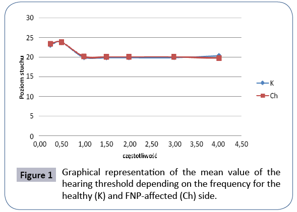
The analysis found that the mean hearing threshold value calculated for selected frequencies was within the normal range for the whole test group, both on the healthy and FNP-affected sides. The t-student test was used to compare the threshold values for selected frequencies between the healthy (K) and affected sides (Ch). No significant difference between the mean hearing threshold for all of the selected frequencies of the FNPaffected and healthy sides was found (Tables 1 and 2 and Figure 1).
| Frequency in kHz | Mean value of the hearing threshold in dB - FNP-affected side | Mean value of hearing threshold in dB - Healthy side | t | p |
|---|---|---|---|---|
| 0.25 | 23.45 ± 3.83 | 23.18 ± 3.77 | 0.38 | 0.708 |
| 0.50 | 23.82 ± 3.84 | 23.82 ± 3.60 | 0.00 | 1.000 |
| 1.00 | 20.18 ± 0.94 | 20.00 ± 0.00 | 1.43 | 0.156 |
| 1.50 | 20.09 ± 0.67 | 20.00 ± 0.00 | 1.00 | 0.320 |
| 2.00 | 20.09 ± 0.67 | 20.00 ± 0.00 | 1.00 | 0.320 |
| 3.00 | 20.09 ± 0.67 | 20.00 ± 0.00 | 1.00 | 0.320 |
| 4.00 | 19.73 ± 1.50 | 20.36 ± 2.70 | 1.53 | 0.129 |
Table 1: Results of the audiometric examination. Comparison of the mean value of the hearing threshold for the healthy and FNP-affected sides.
| Frequency in kHz | Z – non-idiopathic FNP NZ - idiopathic paralysis |
Hearing threshold value | Probability Turkey's test |
|---|---|---|---|
| 0.25 | Z NZ |
23.25 ± 4.06 23.57 ± 3.75 |
0.794 |
| n0.50 | Z NZ |
23.75 ± 3.93 23.86 ± 3.85 |
0.931 |
| 1.00 | Z NZ |
20.25 ± 1.12 20.14 ± 0.85 |
0.723 |
| 1.50 | Z NZ |
20.00 ± 0.00 20.14 ± 0.85 |
0.508 |
| 2.00 | Z NZ |
20.00 ± 0.00 20.14 ± 0.85 |
0.508 |
| 3.00 | Z NZ |
20.00 ± 0.00 20.14 ± 0.85 |
0.508 |
| 4.00 | Z NZ |
19.25 ± 1.83 20.00 ± 1.21 |
0.112 |
Table 2: Comparison of the average hearing threshold value for the individual frequencies between the group of non-idiopathic FNP (Z) and the group of idiopathic FNP (NZ) patients.
The Turkey's range test was used for study groups varying in the number of subjects to compare mean hearing threshold values of patients for which the cause of the disease was known (Z) with patients with idiopathic FNP (NZ).
The analysis did not show significant differences in the mean hearing threshold within the selected frequency range.
Discussion
Evaluation of the hearing organ in patients with peripheral FNP is a very interesting issue. The literature available shows that most of the published results of audiometry of FNP patients are based on electrophysiological methods that focus primarily on the extra-labyrinthal part of the auditory pathway [12,13].
Evaluation with the use of pure-tone audiometry proves to be a very good method for hearing assessment in FNP patients. Results published by numerous authors are contradictory.
In studies in which the tests were performed on a homogenous group of patients with a low average age, without a history of systemic diseases such as diabetes, arteriosclerosis, neoplastic diseases and herpes zoster oticus, the threshold of patients both on the healthy and impaired side was within the norm [10,14,15]. When the studies were conducted among patients with a high mean age, who had a history of other medical diseases, the prevalence of sensorineural hearing loss mainly for high frequencies was significant and ranged from 15 to 50% of the studied group. It should be emphasized that in these cases, hearing loss was bilateral [6,12,16-18].
Many authors emphasize the role of broadly defined polineuropathy also affecting the central nervous system and confirmed in electrophysiological tests in FNP [18,19]. Parameters in the auditory brainstem response (ABR) record that confirm the possibility of auditory pathway injury in patients with FNP are increase of V wave latency, increase of the interval between the I and V waves and reduction in the amplitudes of individual waves. Such results have been obtained in a significant percentage of cases. An additional argument supporting the auditory pathway injury thesis was the normal result of pure-tone audiometry and the presence of the acoustic reflex. An explanation for this phenomenon may be the fact that the neurons of the auditory pathway that lie in the brainstem are closely adjacent to the motor nucleus of the facial nerve [15].
Maurizi et al. performed detailed audiological diagnostics on a group of 30 patients with peripheral FNP. Hearing loss was not confirmed in any patient on the side affected by the palsy with the use of pure-tone audiometry and a lack of the auditory reflex was present in 80% of patients. The researchers also conducted ABR in which a slight increase of the interval between the I and V waves was seen in only two patients, with also presented with normal results of evoked auditory potentials also within the range of middle-latency responses (MLR). Based on their research, they reached a conclusion that FNP is an isolated mononeuropathy and not as it was earlier assumed-a polyneuropathy associated with injury of the CNS.
Hendrix also failed to confirm with the use of ABR any injury of the extranabyrinth part of the auditory pathway, although the lack of the auditory reflex in AI (impendance audiometry) may suggest damage of other pathways in the brain [19]. A common symptom of FNP is reduced tolerance to high intensity tones, which manifests as auditory hypersensitivity, phonophobia and impaired speech (dysacusis). Other symptoms associated with the lack of the auditory reflex are a sudden and difficult to accept increase in auditory sensation. This is disproportionate to the increased intensity of sound sensation (pseudorecruitment) and distortion of speech cognition manifesting in a reduced discrimination in verbal audiometry (rollover) [16,18].
The above mentioned phenomena are typical for labyrinth function impairment. Authors associate some cases of FNP with an extrachochlear injury of the cochlear nerve, thus attributing the etiology to damage of both nerves. However, the only undoubtedly proven mechanism that is able to influence the impairment of the hearing organ in patients with FNP is lack of the auditory reflex [10,16].
Conclusion
The results of subjective pure-tone audiometry showed no evidence of hearing impairment on the FNP-affected side in any of the patients. It also did not show any statistical significant difference between the mean values of the hearing threshold when comparing the affected and non-affected sides.
References
- Mortazavi MM, Latif B, Verma K, Adeeb N, Deep A, et al. (2014) The fallopian canal: A comprehensive review and proposal of a new classification. Childs Nerv Syst 30: 387-395.
- Shoja MM, Oyesiku NM, Griessenauer CJ, Radcliff V, Loukas M, et al. (2014) Anastomoses between lower cranial and upper cervical nerves: A comprehensive review with potential significance during skull base and neck operations. Part I: Trigeminal, facial and vestibulocochlear nerves. Clin Anat 27: 118-130.
- De Seta D, Mancini P, Minni A, Prosperini L, De SE, et al. (2014) Bell's palsy: Symptoms preceding and accompanying the facial paresis. ScientificWorldJournal 14: 801-971.
- Kuhn M, Heman ASE, Jamil A, Shaikh BA, Roehm PC (2011) Sudden sensorineural hearing loss: A review of diagnosis, treatment and prognosis. Trends Amplif 15: 91-105.
- Newadkar UR, Chaudhari L, Khalekar YK (2016) Facial palsy, a disorder belonging to influential neurological dynasty: Review of literature. N Am J Med Sci 8: 263-267.
- Peitersen E (2002) Bell’s palsy: The spontaneous course of 2,500 peripheral facial nerve palsies of different etiologies. Acta Otolaryng 549: 4-30.
- Grønhøj LC, Gyldenløve M, Jønch AE, Charabi B, Tümer Z (2015) A three-generation family with idiopathic facial palsy suggesting an autosomal dominant inheritance with high penetrance. Case Rep Otolaryngol 45: 683-938.
- McNamara R, Doyle J, Mc Kay M, Keenan P, Babl FE (2013) Medium term outcome in Bell's palsy in children. Emerg Med J 30: 444-446.
- Mooney T (2013) Diagnosis and management of patients with Bell's palsy. Nurs Stand 28: 44-49.
- Wormald PJ, Rogers C, Gatehouse S (1995) Speech discrimination in patients with Bell's palsy and a paralyse stapedius muscle. Clin Otolaryngol Allied Sci 20: 59-62.
- Thirumala PD, Ilangovan P, Habeych M, Crammond DJ, Balzer J (2013) Analysis of interpeak latencies of brainstem auditory evoked potential waveforms during microvascular decompression of cranial nerve VII for hemifacial spasm. Neurosurg Focus 34: 20-26.
- Lahin T, Vasama J, Makela JP (2000) Auditory cortical responses in patients with Bell’s Palsy. Acta Otolaryngol 120: 47-50.
- Kaviani M, Jafary AH (1995) Auditory brain-stem response audiometry in patients with Bell’s palsy. Clin Otolaryngol 20: 135-138.
- Maurizi M, Ottaviani F, Almadori G, Falchi M, Paludetti G (1987) Auditory brainstem and middle-latency responses in Bell’s palsy. Audiology 26: 111-116.
- Rosenhall U, Edstrom S, Hanner P, Badr G, Vahlne A (1983) Auditory brain - Stem response abnormalities in patients with Bell’s palsy. Otolaryngol Head and Neck Surg 9: 412-416.
- Candless GA, Schumacher MH (1979) Auditory dysfunction with facial paralysis. Arch Otolaryngol 105: 271-274.
- Rahko T, Karma P (1988) High frequency audiometry in facial paralysis. Acta Otolaryngol 449: 161-163.
- Welkoborsky H, Amedee R, Elkhatieb A, Mann W (1991) Auditory evoked brain-stem responses and auditory disorders in patients with Bell’s palsy. Eur Arch Oto Rhino Laryngol 248: 417-419.
- Hendrix RA, Melnick W (1983) Auditory brainstem response and audiologic tests in idiopathic facial nerve paralysis. Otolaryngol Head Neck Surg 91: 686-690.
Open Access Journals
- Aquaculture & Veterinary Science
- Chemistry & Chemical Sciences
- Clinical Sciences
- Engineering
- General Science
- Genetics & Molecular Biology
- Health Care & Nursing
- Immunology & Microbiology
- Materials Science
- Mathematics & Physics
- Medical Sciences
- Neurology & Psychiatry
- Oncology & Cancer Science
- Pharmaceutical Sciences

