Developmental Sciences from ′Humuncles′ to CRISPR/Cas9
Department of Biomedical and Molecular Sciences, Queen’s University, Kingston, Ontario, Canada
- Corresponding Author:
- Sidra Shafique
Department of Biomedical and Molecular Sciences
Queen’s University, Kingston, Ontario, Canada
Tel: 1-613-533-2727
Fax: 1-613-533-2022
E-mail: s.shafique@queensu.ca
Received Date: September 18, 2018; Accepted Date: October 05, 2018; Published Date: October 10, 2018
Citation: Shafique S (2018) Department of Biomedical and Molecular Sciences, Queen’s University, Kingston, Ontario, Canada. Res J Congenit Disease Vol. 1 No. 3:01.
Copyright: © 2018 Shafique S. This is an open-access article distributed under the terms of the Creative Commons Attribution License, which permits unrestricted use, distribution, and reproduction in any medium, provided the original author and source are credited.
Abstract
Developmental science is a broad term that collectively includes Embryology and Developmental Biology. In the present era, each of these specialty has multiple subspecialties and niches. Here, we are interested to look back in time, the beginning of curiosity and the journey of understanding of reproduction step by step, from sperm in 600 BC to ovum, fertilization, assisted reproductive techniques, in vitro fertilization, inter-species chimera and CRISPR/Cas9 in 2018. The eras of discovery included 600 BC to 1827 (first), 1827 to 1950s (second) and 1950s to present (third). Discoveries and research have always been excited in every era and were highly influenced by the biotechnological advancements. Today, CRISPR/Cas9 is the future of embryo editing with therapeutic potentials and ability to shape the human future. In spite of its prematurity and technical limitations, it is still an active area of research at the cross-points of gene editing, developing inter-species chimeras, treatment of infertility, cure of genetic diseases and organ transplantation.
Keywords
Developmental sciences; History of embryology; Fertilization; Genetic diseases; Organ transplantation
Introduction
Embryology is the branch of developmental biology in which the formation and development of an embryo are studied. Embryonic period of development commences from fertilization and ends at completed eight weeks or 56 days [1]. After 1950s, researchers started using the term ‘Developmental biology’ that referred to the study of cell growth, cell differentiation and the formation of tissues and organs in an organism [2]. Evolution of embryology as a science can be divided broadly over three periods of time depending upon the discoveries regarding sperm, ovum, fertilization, and embryogenesis [3]. The First period extends from the earliest known history until the discovery of ovum in 1827. The Second period covers the discovery of ovum, fertilization, and embryogenesis ending in 1950 while the Third period commences after 1950 as research directed towards the discovery of DNA till date. Each period has distinct milestones of discoveries, eminent scientists and theories that carved the pathways to the development of embryology as a science of the present day. As we go along the history, the effort of the earliest researchers and thinkers needs to be highly admired since they all could not see anything with the naked eye but just visualize the processes of development.
Embryo editing and chimera by CRISPR/CAS9
The sequencing projects such as the Human genome project has been proved to be a stepping stone towards today’s genome engineering technologies development. Today we know the DNA sequences that result in genetic or inherited diseases and we are able to edit these genes through gene editing systems. Genome engineering applications modify genome sequences. Currently, CRISPR/Cas9 is the most advanced biotechnological tool available for gene editing with its vast applications. CRISPR is an abbreviation of Clustered Regularly Interspaced Short Palindromic Repeats. In the CRISPR/Cas9 system where CRISPR is short prokaryotic DNA consisting of repetitive bases and Cas9 is an endonuclease system. The Cas9 consists of “guide RNA” and Cas9 mRNA that are able to find the target DNA to be spliced. Guide RNA helps find the Cas9 the gene to be edited. and a double-stranded DNA break is created followed by the repair along with specific DNA sequence modification [4]. CRISPR/Cas9 is currently being used for gene-editing, interspecies chimera formation, blastocyst complementation system and gene knockout experimental animal production.
To study the human embryology, we need human embryos therefore, the clinically discarded experimental human embryos become available that are abnormal or even if normal, have already been rejected. These embryos have the potential to hatch into blastocysts. The zygotes may contain two pronuclei (normal) or zero, one, three, four or more pronuclei (abnormal). The blastocyst development rate is 63.8% for two cell embryos while 22.6% of two pronuclei zygotes. These clinically discarded in vitro fertilization material is used for the experiments of CRISPR/Cas9 gene editing, gene function studies and mutations [5]. In a recent report, Kang, et al. [6] injected donor oligo-1 (99 bp) or oligo-2 (99 bp) (100 ng/μl) or dsDonor (1 kb) mixed with Cas9 mRNA (100 ng/μl) and gRNA (50 ng/μl) and successfully introduced CCR5Δ32 allele into early human 3PN embryos using CRISPR/Cas9 system. Interspecies chimerism with CRISPR/Cas9- mediated zygote genome editing is being used to produce the species-specific organs with the prospect of future organ transplantation. Blastocyst complementation is the method in which the host blastocysts are obtained and gene editing is done using the CRISPR/Cas9 system [7].
The first era of the developmental sciences: 600 BC to 1827
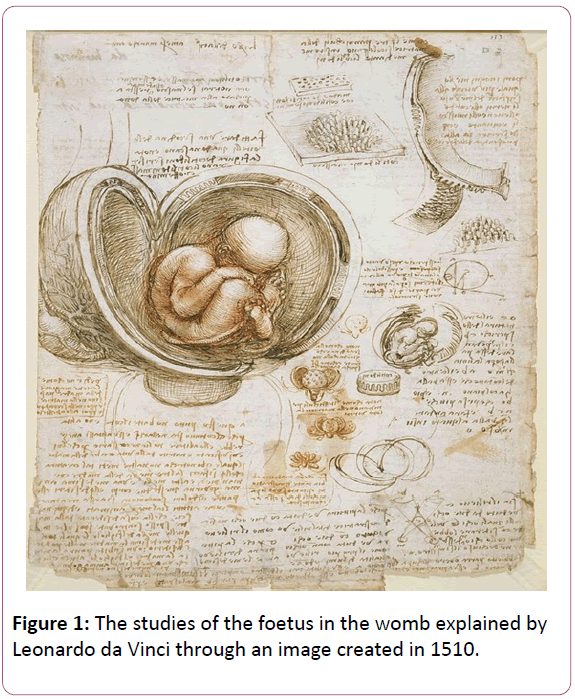
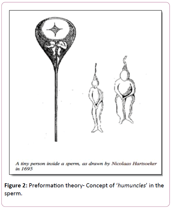
The First era started about 600 BC and the earliest thinker known as Pythagoras (about 600 BC), who originated “spermism”, the doctrine. According to this doctrine, the father contributes essential characteristics to the developing offspring and the mother just lends the womb (Figure 1) [8]. Hippocrates (460-370 BC) suggested that there is a contribution of both parents to generate a “seed”. His text proposed that developing offspring grows inside a membrane and nourished by mother’s blood. Hippocrates was the first to give the idea of opening and examining the hen eggs on a daily basis to look for the growth of the offspring [9]. The popular theory at that time was ‘Preformationism’ which states that organisms develop from miniature versions of themselves (Figure 2). These were called “homunculi” for humans and “animalcules” for the animals. Homunculi and animalcule were supposed to be present in the male secretions and placed in the female womb just to grow in size [10].
Aristotle (384-322 BC) studied chick embryo systematically and produced accurate drawings of the embryo growth till the chick hatched. He proposed the theory of Epigenesis also known as neoformation. It states that undifferentiated mass passes through a series of steps during which new parts are added leading to gradual development of an organism [11]. Aristotle was first to describe the umbilical cord, development of the beating heart, blood vessels and blood as the source of nourishment for the growing embryo. His theory of epigenesis favored the emergence of an organism from a formless egg. Later on, the advent of the microscope confirmed that embryo takes shape in a progressive series of steps along the growth from a single egg [12]. Followed by the Greek era, Galen (129-210) studied the female reproductive system including the uterus, “female testes” and oviducts. Vesalius, an Italian anatomist and his pupil Fallopius provided the detailed description of the fallopian tubes, which were named after him [13]. Fabricius, the intelligent student of Fallopius, was an Italian anatomist. He studied the formation of hen’s egg in the organ that he named ovarium, presently known as ovary [14]. His English student, William Harvey, studied deer and other mammals, after mating, by dissection. He wrote a book ‘On the Generation of Animals’ in 1651 and asserted for ex ovo Omnia, all animals come from eggs. Harvey is considered the father of ovist preformationism.
Although he was still epigenesist about the process of development yet he challenged most of the ideas proposed by Aristotle and revolving around more since fifteen hundred years [15].
Stensen, introduced the term ‘ovary’ and De Graaf, confirmed the presence of eggs in the ovary. The eggs were named after De Graaf as the Graafian Follicles [16]. In 1677, a Dutch microscopist, Leeuwenhoek observed the large number of moving organisms in his own semen, which he named as “spermatic worms” or animalcules. Later on, he demonstrated that semen having living animalcules was present inside the uterus of dogs after mating [17]. It was thought that animalcules contain all the components of the embryo and uterus is only the site of incubation [15]. Victor Haller was convinced with de Graaf’s follicles and proposed that conception occurs in the ovary. According to his theory, follicle discharges the preformed embryo in the oviduct and oviduct transfers it into the uterus where it grows further [18].
Lazzaro Spallanzani, an Italian biologist in 1785, did experiments on frogs and concluded that semen must come in contact with the ovum to create an embryo [19]. A Swiss physiologist, Jean-Louis, and a French chemist Dumas conducted experiments in 1824. They were the leading scientists who actually looked into the Graafian follicle and embryo under the microscope. They observed that even after twelve days of conception, the embryo was smaller in size than the Graafian follicle protruding from the surface of the ovary. Therefore, they discarded the theory and postulated that it must not be an embryo that ruptured in the oviduct as they were grossly different in size [16].
The second era of the developmental sciences: 1827 to 1950s
In the Second era, Karl Ernst von Baer, a Russian biologist, finally established in 1827 that there is a true ovum of mammals, “ovulum” [20]. By 1834, as described by the Bernhardt, the polish biologist, ovulum was known as “the germinal vesicle”. Meanwhile, Charles Darwin conceived the theory of natural selection in 1838. He is considered one of the pioneers of modern embryology [21]. After the cell theory emerged, by Schwann and Schleiden, the ovum was admitted to be a cell and structural unit. In 1882, Walter Flemming described the chromosomes, meiosis, and mitosis. This led to the cascade of discoveries, thus the ovum maturation was established by 1884 [22]. Walter Heape was the first researcher who successfully recovered the eggs from the rabbit [23]. He also transferred fertilized eggs, in utero, to different animals. Sobotta provided the detailed account of mouse egg maturation, fertilization, and cleavage with histological evidence. An even big breakthrough was in vitro culture and development of the fertilized rabbit eggs by Lewis and Gregory. They were able to record a video, for the first time, of the cleavage divisions and provided the visual evidence of embryonic development.
Lewis and Wright in 1935 photographed the cleavage stages of the mouse [24]. Later on, Whitten extended this work and showed that eight-cell embryos could be cultured to blastocysts. It was followed by in vitro development of late two-cell stage embryos. The ability of in vitro embryo culture improved through late stages of development to the earlier stages as research advanced in embryology. The most important difficulty for in vitro culture, clearly pointed out by the promoter of “causal embryology” the Van Benden around 1920, was lack of effective tissue culture techniques [25]. The pioneering work regarding in vitro fertilization of human eggs was reported by Rock, et al. [26]. About eight hundred ova were collected from women, through various surgical procedures, in the late follicular phase. One hundred and thirty-eight ova were exposed to sperms in different culture conditions. This work led to the discovery of capacitation of sperm and pioneered clinical in vitro fertilization. There is an exhaustive list of experimentation, followed by Rock and Menkin work, that was focused on improvements in embryo culture; both for experimentation and clinical purposes for infertility treatment.
The third era of the developmental sciences: the 1950s-to-date
After the 1950s, the embryology entered in the Third era that is marked by successes in multiple directions. Watson, et al. [27] discovered the double helical structure of the DNA molecule. This discovery now opened the doors of research towards genetics, cell biology, and pre-implantation genetic diagnosis. Brinster [28] reported comprehensive research work on the essential ingredients required for the normal preimplantation embryonic development. Immense laboratory work was done regarding the optimum pH, osmolarity, amino acid requirements and culture volume as well as physical support that helped successful embryo development. It led to successful attempt of in vitro fertilization and Lousie, the first IVF female baby [29], was born in England by Dr. Edwards and Dr. Steptoe. This milestone opened the door of successful IVF conceptions and live births all over the world. In Canada, first IVF baby was delivered by Gomel and Poland in December 1983.
Clinical embryology has distinctly been separated from experimental embryology. Contemporary directions in clinical embryology include Pre-implantation Genetic Diagnosis (PGD), improvements in assisted reproductive techniques for better success rates in live births and better maternal receptivity for the implanted embryo. In present days, the cutting edge research in laboratories includes stem cell studies, cloning and genetic control over developmental processes. Teratology, a study of developmental anomalies, flourished after successful in vitro fertilization and ability to observe the embryo development.
Conclusion
The insight that originated about 2500 years ago in Pre-Greek era evolved through so many diverse cultures and minds. What embryology has achieved now, is predominantly the outcome of research work carried out in the last 200 years. Now embryology is not only one discipline, rather it possesses many subdisciplines of its own. The religious, cultural and ethical conditions over all times were challenging for the scientists working in developmental sciences. Nonetheless, it is a true basic science for every other science as embryology makes the foundation. The efforts of all, who contributed to its development as a science, will be remembered both for inspiration and their devotion towards research.
References
- Necatholic.org/wp-content/uploads/.../Second-Look-Fetal-Development.pdf
- Developmental biology. ScienceDaily.
- Carlson, Bruce M, Kantaputra, Piranit N (2014) 4 molecular basis for embryonic development. Human Embryology and Developmental Biology (5th edn.).
- Hsu PD, Lander ES, Zhang F (2014) Development and applications of CRISPR-Cas9 for genome engineering. Cell 157: 1262-1278.
- Yao G, Xu J, Xin Z, Niu W, Shi S, et al. (2016) Developmental potential of clinically discarded human embryos and associated chromosomal analysis. Sci Rep 6: 23995.
- Kang X, He W, Huang Y, Yu Q, Chen Y (2016) Introducing precise genetic modifications into human 3PN embryos by CRISPR / Cas-mediated genome editing. J Assist Reprod Genet 33: 581-588.
- Wu J, Platero-Luengo A, Sakurai M, Sugawara A, Gil MA, et al. (2017) Interspecies chimerism with mammalian pluripotent stem cells. Cell 168: 473-486.
- An Johnston (2000) In: And still we evolve: A handbook on the history of modern science. Section Five: Heredity and Modern Genetics.
- Cole FJ (1930) Early theories of sexual generation. Clarendon Press, Oxford.
- Maienschein J (2005) Epigenesis and preformationism. Stanford encyclopedia of philosophy (Fall 2008 Edition), Edward N. Zalta (ed.).
- Magner Lois (2002) A history of the life sciences. New York: Marcel Dekker, Inc, pp: 1-154.
- Lopata A (2009) History of the egg in embryology. J Mamm Ova Res 26: 2-9.
- Mortazavi MM, Adeeb N, Latif B, Watanabe K, Deep A, et al. (2013) Gabriele Fallopio (1523-1562) and his contributions to the development of medicine and anatomy. Childs Nerv Syst 29: 877-880.
- Smith SB, Macchi V, Parenti A, de Caro R (2004) Hieronymous fabricius from acquapendente (1533-1619). Clinical Anatomy 17: 540-543.
- Correia CP (1997) The ovary of eve: egg and sperm preformation. Chicago: University of Chicago Press.
- Corner GW (1933) The discovery of the mammalian ovum. In: Lectures on the History of Medicine, WB Saunders Company, Philadelphia, pp: 401-426.
- Friedman DM (2001) A mind of its own: A cultural history of the penis. New York: The Free Press.
- Austin CR (1961) The mammalian egg. The mammalian egg. pp: 12-13.
- Gasking EB (1967) Investigations into generation. 1651-1828, pp: 130-136.
- Betteridge KJ (1981) An historical look at embryo transfer. Reproduction. J Reprod Fertil 62: 1-13.
- Beddall BG (1968) Wallace, Darwin, and the theory of natural selection: a study in the development of ideas and attitudes. J Hist Biol 1: 261-323.
- Sander K (1991) William Roux and his program for developmental biology. Roux's Arch Dev Biol 200: 1-3.
- Heape W (1890) Preliminary note on the transplantation and growth of mammalian ova within a uterine foster-mother. Proceedings of the Royal Society of London 48: 457-458.
- Lewis WH, Wright ES (1935) On the development of mouse. Contrib Embryol Carnegie Inst Wash 25: 113-146.
- Whitten WK (1956) Culture of tubal mouse ova. Nature 176: 96.
- Rock J, Menkin MF (1944) In vitro fertilization and cleavage of human ovarian eggs. Science 100: 105-107.
- Watson JD, Crick FHC (1953) A structure for deoxyribose nucleic acid. Nature 171: 737-738.
- Brinster RL (1965) Studies on the development of mouse embryos in vitro. J Reprod Fertil 10: 227-240.
- World's first IVF baby marks 30th birthday. Agence France-Presse, 23 July 2008.
Open Access Journals
- Aquaculture & Veterinary Science
- Chemistry & Chemical Sciences
- Clinical Sciences
- Engineering
- General Science
- Genetics & Molecular Biology
- Health Care & Nursing
- Immunology & Microbiology
- Materials Science
- Mathematics & Physics
- Medical Sciences
- Neurology & Psychiatry
- Oncology & Cancer Science
- Pharmaceutical Sciences


