Case Report on Pleural Aspergillosis in 52 Year Female Cancer Patient
1The University of the West Indies (UWI), St. Augustine, Trinidad and Tobago
2North Central Regional Health Authority, Eric Williams Medical Sciences Complex, Champ Fleurs, Trinidad and Tobago
- *Corresponding Author:
- Umakanthan S, MBBS
MD-Consultant, Pathologist, Lecturer
The University of the West Indies
Trinidad and Tobago
Tel: 1-868-645-2645
E-mail: srikanth.umakanthan@sta.uwi.edu
Received Date: August 12, 2017; Accepted Date: August 30, 2017; Published Date: September 11, 2017
Citation: Umakanthan S, Bukelo MM (2017) Case Report on Pleural Aspergillosis in 52 Year Female Cancer Patient. J Oncopathol Clin Res. 1:2.
Abstract
Pleural Aspergillosis is a rare entity, with most of the cases occurring on a background of lung disease or surgery. We report a case of a 52-year-female who developed pleural Aspergillosis. Patient presented with dyspnoea and weight loss of 6 weeks duration. A chest radiograph revealed features of the pleural effusion with lung collapse. Histopathology evaluation confirmed Aspergillus. We report this case in view of the extreme rarity of pleural Aspergillosis occurring in a setting of previous breast carcinoma.
Keywords
Pleural Aspergillosis; History of dyspnoea; Fungal infection
Case Report
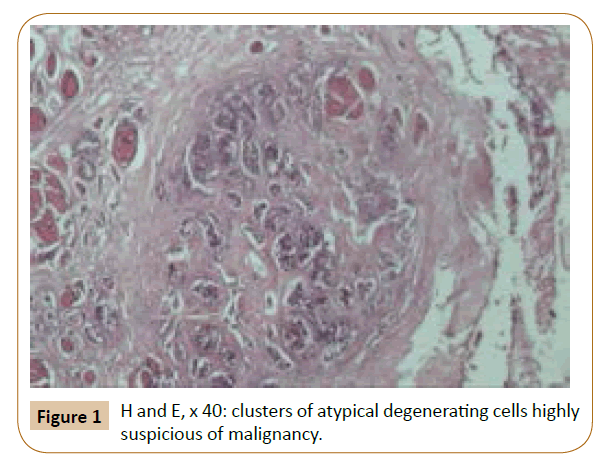
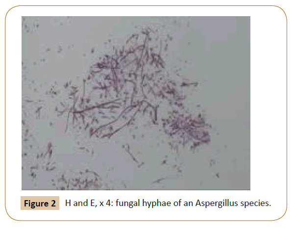
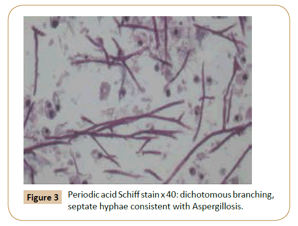
A 52 year female presented with an 8 weeks history of dyspnoea and weight loss. There was a previous history of breast carcinoma. Chest roentgenograms showed pleural effusion with lung collapse. Decorticated right pleural tissue was submitted for histopathological evaluation. On gross pathology finding six fragments of tan friable tissue were noted. Histology revealed parietal pleural tissue with area of atypical degenerating cells, highly suspicious of malignancy along with branching septate hyphae suggestive of fungal infection. Further, Periodic acid Schiff Stain showed numerous dichotomous branching, septate hyphae consistent with Aspergillosis. The type of malignancy could not be identified.
Discussion
Aspergillus is a ubiquitous fungus belonging to the family Aspergillacea and was first described by Cleland in 1924 and as an opportunistic infection in 1953 [1]. The route of infection is through respiratory and gastrointestinal tracts and usually seen in immunocompromised patients. Invasive aspergillosis is seen in a healthy or mildly immunocompromised host. Recently, the genus Aspergillus is subdivided into 8 subgenera and 22 sections [2]. The spectrum of diseases caused by Aspergillus species varies from superficial cutaneous to invasive and systemic infections.
Clinical presentation in most cases is asymptomatic with an underlying chronic disorder such as diabetes. Symptoms become apparent as the disease progresses over months and presents with diplopia, nasal stuffiness in the absence of fever [3]. Histopathologically, hyphal forms can be seen usually in the area of necrosis. They appear as septate hyphae, with branching at 45° angles and are about 2-4 mm in diameter. This fungus differentiated from mucormycosis was broader non-septate hyphae with dichomatous branching at 90° angles are observed as in our case [4]. Conventional staining by haematoxylin and eosin can be confirmed by special stains such as periodic acid- Schiff (PAS), Gomori’s methenamine silver (GMS), and immunocytochemistry. In our case PAS was performed for confirmation [5].
Pleural Aspergillosis is a rare entity which occurs in a setting of tuberculosis due to breakdown of pulmonary immune mechanism and also in established empyema and bronchopleural fistula [6]. In our case, pleural infection developed together with a malignancy of unknown origin in a setting of previous breast carcinoma which makes it unique (Figures 1-3).
Figure 1: H and E, x 40: clusters of atypical degenerating cells highly suspicious of malignancy.
Microbiologists, pathologists, and clinicians need to be aware of the limitations of tissue diagnosis, the pitfalls of morphological diagnosis, and the tests that can be performed with tissue and other samples to make organism-specific diagnoses especially in Aspergillus infection.
References
- Bhatnagar T, Bhatnagar AK (2014) Pleural Aspergillosis in an otherwise healthy individual. Lung India: Official Organ of Indian Chest Society 31: 155-157.
- Tamgadge AP, Mengi R, Tamgadge S, Bhalerao SS (2012) Chronic invasive aspergillosis of paranasal sinuses: A case report with review of literature. J Oral Maxillofac Pathol 16: 460-464.
- Sharma OP, Chwogule R (1998) Many faces of pulmonary aspergillosis. Eur Respir J 12: 705-715.
- Buzina W (2013) Aspergillus - classification and antifungal susceptibilities. Curr Pharm Des 19: 3615-3628.
- Karthik RK, Sudarsanam TD (2009) An unusual cause of empyema thoracis. Indian J Med Sci 63: 30-32.
- Krakówka P, Rowiñska E, Halweg H (1970) Infection of the pleura by Aspergillus fumigatus. Thorax 25: 245-253.
Open Access Journals
- Aquaculture & Veterinary Science
- Chemistry & Chemical Sciences
- Clinical Sciences
- Engineering
- General Science
- Genetics & Molecular Biology
- Health Care & Nursing
- Immunology & Microbiology
- Materials Science
- Mathematics & Physics
- Medical Sciences
- Neurology & Psychiatry
- Oncology & Cancer Science
- Pharmaceutical Sciences


