


 26 / 65

Page 46
May 24-25, 2018
London, UK
Vascular Surgery 2018
3
rd
Edition of World Congress & Exhibition on
Vascular Surgery
Journal of Vascular and Endovascular Therapy
ISSN: 2573-4482
Objective:
To study the relevance between the incidence of
residual stenosis and carotid artery stent (CAS) characteristics
by color duplex flow imaging (CDFI).
Methods:
Five hundred and seventy two cases (576 stents, open
or closed-cell stents) who underwent CAS from January 2013 to
December 2015 were retrospectively enrolled in this study. The
location of carotid stenosis (common carotid artery or internal
carotid artery), characteristics of plaques (regular morphology or
not; with calcification or not), the length of stent, types of carotid
stent (closed or open cell), rate of stent expansion (ratio of radial
expansion and axial expansion) were detected one month before
and one week after stenting by CDFI. Residual stenosis is defined
as the stenosis rate is equal to or greater than 30% by DSA
immediately after stenting.
Results:
All of 576 stents, the incidence of residual stenosis was
significantly higher in group of closed loop stent (28.3%, 46/163)
than in group of open loop stent (20.4%, 84/413) (2=4.15, P=0.04).
There were positive correlation between the occurrence rate of
residual stenosis and closed-cell stent (odd ratios, 1.54; 95%
confidence interval, 1.02-2.23) and negative correlation with the
radial expansion rate (odd ratios, 0.02; 95% confidence interval,
0.01-0.06). The location of carotid stenosis and the lengths of
stents were not affecting the incidence of residual stenosis.
Irregularly shaped plaques (odd ratios, 9.72; 95% confidence
interval, 5.65-16.76) and the plaques with calcification (odd ratios,
5.21; 95% confidence interval, 3.32-8.17) were the independent
risk factors of residual stenosis after CAS.
Conclusions:
This study suggests that choosing a more adaptable
stent based on the types of stents and the characteristics of
plaques and trying to increase the radial expansion of stenting
may further decrease incidence rates of residual stenosis.
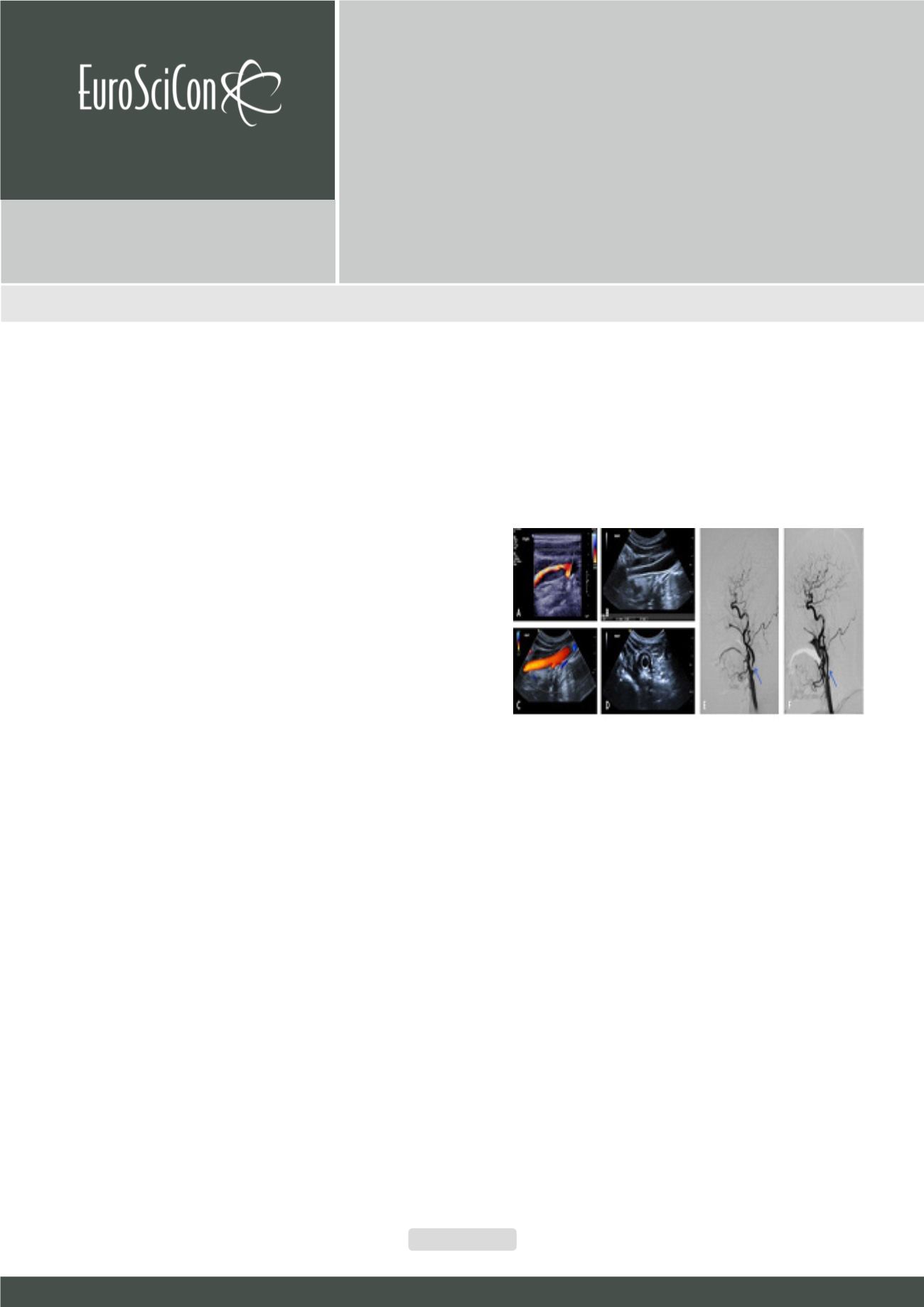
Figure 1:
A: CDFI showed a severe (70-99%) stenosis at the proximal internal
carotid artery; B & D: 2-D images of longitude view and cross-section view
showed residual stenosis after CAS; C: CDFI showed the blood flowwithin the
stent was smooth; E: DSA before CAS showed a severe stenosis at the proxi-
mal internal carotid artery (blue arrow); F. DSA immediately after CAS showed
a residual stenosis of the stent (blue arrow).
Recent Publications
1. Csobay Novak C, Barany T, Zima E et al. (2015) Role
of stent selection in the incidence of persisting
hemodynamic depression after carotid artery stenting. J
Endovasc Ther 22(1):122-129.
2. Fujii K, Carlier S G, Mintz G S, et al. (2005) Stent
underexpansion and residual reference segment
stenosis are related to stent thrombosis after sirolimus-
eluting stent implantation: an intravascular ultrasound
study. J Am CollCardiol 45(7):995-998.
3. Tsutsumi M, Kodama T, Aikawa H, et al. (2010)
Fragmentation of calcified plaque after carotid artery
stenting in heavily calcified circumferential stenosis.
Affecting factors of residual stenosis after carotid artery
stenting
Lingyun Jia, Yang Hua, Yunlu Tao, Liqun Jiao, Lili Wang, Beibei Liu
and
Weihong Hou
Xuanwu Hospital of Capital Medical University, China
Lingyun Jia et al., J Vasc Endovasc Therapy 2018, Volume 3
DOI: 10.21767/2573-4482-C1-002