Folic Acid Improves Vascular Endothelial Dysfunction in Children with Type-1 Diabetes Mellitus
Suzan Omar Mousa1*, Samir Tamer Abd-Allah1, Mohamed Abdel-Razek Abdel-Hakem2 and Sara Gamal Hares1
1Department of Pediatrics, Minia University, Egypt
2Department of Clinical Pathology, Minia University, Egypt
- *Corresponding Author:
- Suzan Omar Mousa
Department of Pediatrics
Faculty of Medicine, Minia University
El-Minya, Egypt
Tel: 201006163560
Fax: 20862337634
E-mail: suzanmousa@mu.edu.eg
Received Date: Aug 10, 2017; Accepted Date: Oct 30, 2017; Published Date: Nov 10, 2017
Citation: Mousa SO, Abd-Allah ST, Abdel-Hakem MA, Hares SG (2017) Folic Acid Improves Vascular Endothelial Dysfunction in Children with Type-1 Diabetes Mellitus. Endocrinol Metab Vol. 1 No. 1:105.
Copyright: © 2017 Mousa SO, et al. This is an open-access article distributed under the terms of the Creative Commons Attribution License, which permits unrestricted use, distribution and reproduction in any medium, provided the original author and source are credited.
Abstract
Background: Diabetic endothelial dysfunction stimulates release of inflammatory factors such as von Willebrand factor (vWF) and vascular cell adhesion molecule-1 (VCAM-1) and also results in microalbuminuria. Folic acid had shown to improve endothelial dysfunction.
Objective: To evaluate the effects of folic acid supplementation on VCAM-1, vWF and microalbuminuria in children with type-1 diabetes mellitus. Methods: Our study was conducted upon 30 children with type 1 diabetes mellitus (aged between 8-15 years). They received oral folic acid in a dose of 5mg daily for 3 months. Blood samples were taken before and after folic acid supplementation to evaluate glycosated hemoglobin (HbA1c), vWF, VCAM-1, and albumin to creatinine ratio (A/C ratio).
Results: VCAM-1 and A/C ratio levels were decreased significantly after folic acid administration in the two groups (p<0.001 for each). vWF decreased after folic acid supplementation, but this was of statistical insignificance. On the other hand, HbA1c did not significantly change after folic acid administration (p>0.05).
Conclusion: Folic acid improves endothelial function in diabetic children measured by VCAM-1 and A/C ratio. However, it did not significantly decrease vWF which may need longer periods of folic acid administration.
Keywords
Folic acid; VCAM-1; vWF; Microalbuminuria; Endothelial dysfunction; Diabetes; Children
Introduction
Type-1 Diabetes Mellitus is considered as the second most common chronic disease in children. It is a multifactorial disease with high rates of morbidity and mortality due to microvascular or macrovascular complications [1]. The microvascular complications in diabetes encompass long-term complications affecting small blood vessels including diabetic retinopathy, nephropathy and neuropathy. The macrovascular complications include the diseases of large blood vessels throughout the body including coronary and peripheral arteries leading to cardiovascular and cerebrovascular diseases and stroke [2]. Endothelial dysfunction is the main factor in the progression of vascular complications which occur before the clinical manifestations [3].
Endothelial cells produce specific adhesion molecules, such as E-selectin, intracellular adhesion molecule (ICAM) and vascular cell adhesion molecule (VCAM), for the regulation of cell adhesion and permeability [4]. The intact endothelium expresses low levels of these adhesion molecules, and upon activation, over-expression of these molecules has been reported, which play a role in maintaining endothelial barrier integrity [5]. Also, Endothelial cells have major roles in regulating hemostatic balance, preventing the activation of thrombin and inhibiting platelet adhesion, thereby mediating anticoagulant activity [6]. Upon development of endothelial dysfunction, there is increase in the release of endothelial products, such as von Willebrand factor (vWF), angiopoietin-2 and P-selection, which are involved in the modulation of inflammatory response [7].
Microalbuminuria has been considered an expression of endothelial dysfunction. It is a disorder of the capillary wall in the glomerulus with trans-capillary escape of albumin [8]. In diabetes, endothelial dysfunction has been correlated with microalbuminuria [9] and may precede its development [10].
Folic acid supplementation had shown to improve endothelial dysfunction. The mechanism underlying the improvement in endothelial function remains controversial [11]. As, folic acid increases nitric oxide (NO) bioavailability and decreases oxidative stress, which improves endothelial progenitor cell dysfunction in diabetic patients, through improving endothelial nitric oxide synthase (eNOS) function by a number of suggested mechanisms, such as: 1) Stabilizing tetrahydrobiopterin by preventing its oxidation and stimulating its regeneration from the oxidized dihydrobiopterin 2) Facilitating the binding of tetrahydrobiopterin to eNOS 3) 5-methyltetrahydrofolate may mimic tetrahydrobiopterin at its receptor site on eNOS or may facilitate the electron transfer to produce NO [11].
Our aim was to evaluate the effect of folic acid supplementation on endothelial function in diabetic children. We assessed: urinary albumin/creatinine ratio (A/C ratio) for development of microalbuminuria, regulation of platelet adhesion and aggregation (vWF), leucocyte adhesion (VCAM-1) [12]. The rationale for using these endothelial dysfunction marker proteins is that they reflect endothelial damage.
Material and Methods
Subjects
This prospective cohort study was conducted on 35 diabetic children who had regular follow up in the Pediatric Endocrinology Outpatient Clinic, Minia University Children Hospital. We excluded from our study children who had diabetes for less than 2 years, had any systemic diseases other than diabetes, suffered from DKA or hypoglycemia before blood sampling by 2 weeks or refused to participate in the study.
The study was explained in detail to the parents or legal guardians of the participant children and written consents were taken from them. The study was designed respecting the expected ethical aspects. It was performed according to the Declaration of Helsinki 1975, as revised in 2008 and approved by the Institutional Review Board and Medical Ethics Committee of Minia University Hospital.
All included children were given oral folic acid supplementation in a dose of 5 mg daily for 3 months with regular follow up of the patients checking their compliance. Five patients dropped out due to non-compliance.
Methods
All included children had undergone the following at start of the study and after 3 months of folic acid therapy:
6 ml of morning venous blood after overnight fasting was withdrawn.
2 mL was collected in tubes, left to clot for 30 minutes then the sera were separated from the cells using centrifuge at 1000 xg for 15 minutes and stored at temperature -20°C or less until VCAM-1 assay was done. VCAM-1 measured by Quantikine Human sVCAM-1 immunoassay (ELISA) kit (Minneapolis, United States of America).
2 ml was collected in tube containing sodium citrate as an anticoagulant, centrifuged immediately after collection to separate plasma from cells and then stored at -70°C until von Willebrand facror antigen assay was done. Von Willebrand antigen was measured by (ELISA) kit (Corgenix, United States of America).
2 ml was collected in tubes containing EDTA as anticoagulant for Glycosylated Hemoglobin HbAıc assay, which was measured by Stanbio glycohemoglobin procedure for quantitative colorimetric determination of glycohemoglobin (Stanbio Laboratory, Boerne, Texas).
Urine samples were collected in a clean container under complete aseptic conditions to assess albumin to creatinine ratio (A//C ratio) as an indicator of microalbuminuria. Albumin in urine was measured by turbidmetry method using sulfosalicylic acid reagent (3:1) and creatinine was measured by Mindrray BS 300 chemical analyser.
Statistical methods
The collected data were statistically analyzed using statistical package for social sciences (SPSS) program for windows version 20. Quantitative results were presented as mean ± standard deviation (SD) while qualitative data were presented by frequency distribution as percentage (%). Chi square test (X2) was utilized for analysis of qualitative data, and Student (t) test for analysis of quantitative variables. Correlations were performed by using Pearson’s and Spearman’s correlation coefficient (r). The improvement in the endothelial function markers were calculated by their values before folic acid supplementation-their values after folic acid supplementation. Receiver operating characteristic (ROC) curve analysis was performed using MedCalc_version 12.1.4.0. to determine: the optimal cut-off values and the diagnostic performance of the variable, the diagnostic sensitivity and specificity, and comparison of sensitivity and specificity for VCAM-1 and A/C ratio after folic acid supplementation. p-value if less than 0.05 was considered as a cut off for significance.
Results
Thirty diabetic children were included in our study. Their ages ranged between 8 and 15 years with a mean of 12.2 ± 1.9 years, 11 (36.7%) of them were males while 19 (63.3%) were females, their mean duration of diabetes was 6.7 ± 1.9 years. Family history of diabetes was positive in 14 children (46.7%).
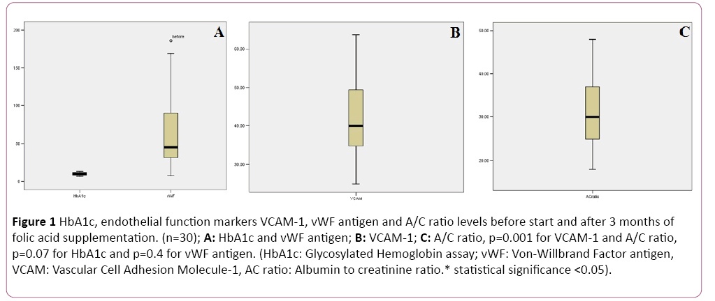
We found significant decrease in VCAM-1 and A/C ratio levels after folic acid supplementation (p<0.001). VCAM-1 and A/C ratio levels before folic acid supplementation were 45.6 ± 9.4 ng/ml and 33.1 ± 8.2 mg/g respectively. While their levels after folic acid supplementation were 37.9 ± 7.5 ng/ml and 28.3 ± 5.8 mg/g respectively. On the other hand, HbA1c and vWF did not show statistically significant changes (p>0.05). As, their levels before folic acid supplementation were 10.3 ± 1.9% and 65.7 ± 47.2% respectively. While their levels after folic acid supplementation were 10.07 ± 1.9% and 56.9 ± 38.4% respectively (Figure 1).
Figure 1: HbA1c, endothelial function markers VCAM-1, vWF antigen and A/C ratio levels before start and after 3 months of folic acid supplementation. (n=30); A: HbA1c and vWF antigen; B: VCAM-1; C: A/C ratio, p=0.001 for VCAM-1 and A/C ratio, p=0.07 for HbA1c and p=0.4 for vWF antigen. (HbA1c: Glycosylated Hemoglobin assay; vWF: Von-Willbrand Factor antigen, VCAM: Vascular Cell Adhesion Molecule-1, AC ratio: Albumin to creatinine ratio.* statistical significance <0.05).
Pearson’s and Spearman correlation tests were performed to study the association of the improvement in VCAM-1, VWF and A/C ratio after folic acid supplementation with the demographic, clinical and HbA1c level. None of the studied markers showed significant correlations with age, family history, sex, duration of diabetes or HbA1c level (p>0.05).
Validity tests were used to demonstrate the optimal cut-off values of VCAM-1 and A/C ratio. VCAM-1, at a cutoff value of ≤ 37.5 ng\ml, was more sensitive (60%) and specific (76.6%) with higher PPV (68.2%) and NPV (63.6%) than A/C ratio to detect the improvement in endothelial function after folic acid supplementation in diabetic children (Table 1).
Table 1 Validity tests of VCAM-1 and A\C ratio in predicting endothelial dysfunction after folic acid supplementation.
| Variables | Cut-off | Sensitivity % | Specificity % | PPV % | NPV % |
|---|---|---|---|---|---|
| VCAM-1 | ≤ 37.5 ng/ml | 60 | 76.6 | 68.2 | 63.6 |
| A\C ratio | ≤ 29 mg/g | 50 | 70 | 66.7 | 60.5 |
VCAM-1: Vascular Cell Adhesion Molecule-1; A/C ratio: Albumin to creatinine ratio; PPV: positive predictive value; NPV: negative predictive value
Discussion
In this study after 3 months of folic acid supplementation, there was significant improvement in endothelial dysfunction represented by significant decrease in VCAM-1 and A/C ratio. The results of the present study may be attributed to the antioxidant properties of folic acid through which it may improve endothelial progenitor cell function [13]. This was in agreement with Alian et al. in 2012 who found that folic acid supplementation improved endothelial dysfunction, as VCAM-1 and microalbuminuria decreased significantly in the studied group [14]. While, a more recent study, by Schneider et al. in 2014, demonstrated that folic acid fails to improve endothelial dysfunction in diabetic nephropathy patients [15]. This contradiction may be attributed to that Schneider’s study was carried out on type 2 diabetic patients, and oral antioxidant treatment was found to improve endothelial dysfunction in type 1 more than type 2 diabetic patients [16].
Alain’s study in 2012 did not find serum vWF to change significantly after folic acid supplementation in a dose of 5mg daily for 2 months [14]. In our study, we gave the same folic acid dose but for 3 months. vWF decreased in our study after folic acid supplementation, but this decrease did not reach statistical significance. Surprisingly, Mierzecki et al. in 2012 found a significant decrease in vWF concentrations in atherosclerotic patients after low-dose folic acid supplementation (0.4 mg daily) for 3 months [17]. This may be attributed to the fact that low dose of folic decrease homocysteine level. Homocysteine inhibits vWF processing and secretion by preventing its transport from the endoplasmic reticulum [18]. High dose of folic acid improves endothelial function, as we mentioned before, through improving eNOS function mainly, and this occurs before changing homocysteine level [19]. So, vWF antigen decreased in our study after folic acid supplementation. Further prolonging the periods of folic acid administration may be necessary to achieve a statistical significant decrease.
HbA1c did not show significant difference before and after folic acid supplementation. This alleviate the role of hyperglycemia on the studied markers. Our results are in accordance with many studies that reported the trend of folic acid supplementation to be associated with better glycemic control, but this control was with insignificant effect on HbA1c levels [14,20]. However, Pena and his study group got two contradicting results regarding the relation of folic acid supplementation and HbA1c level [21,22]. They explained this contradiction by the effects of study participation on patients’ motivation which lead to the significant improvement in HbA1c level [21].
Although, a study by Ebbing et al. in 2009 reported an increased cancer incidence and mortality, especially lung cancer, when folate was co-administrated with vitamin B12 supplementation [23]. But, they did not study the effect of family history of cancer, environmental and occupational factors on their results. Besides, other studies have demonstrated no associations between intakes of folate or folic acid and lung cancer risk [24,25].
VCAM-1, at a cutoff value of ≤ 37.5 ng/ml, was more sensitive (60%) and specific (76.6%) than microalbuminuria measured by A\C ratio, to detect the improvement in endothelial function after folic acid supplementation in diabetic children. VCAM-1 is known to be a dynamic surrogate marker for the effectiveness of therapeutic interventions in diabetic patients with microalbuminuria [26]. Moreover, it adds additional information on cardiovascular risk [27].
Conclusion
Folic acid supplementation improved endothelial dysfunction in children with type-1 diabetes mellitus, and we recommend that all diabetic children should receive oral folic acid supplementation as a part of their chronic therapy. Our study has several limitations, for example, serum homocysteine and folic acid level were not feasible to be assessed. Longer periods of follow up with larger sample size might have been more informative regarding the effect of folic acid on vWF and HbA1c.
Conflict of Interests
All authors declare that they have no conflicts of interests.
Financial Support
No financial support received.
References
- Ceriello A, Esposito K, Ihnat M, Thorpe J, Giugliano D (2009) Long-term glycemic control influences the long-lasting effect of hyperglycemia on endothelial function in type-1 diabetes. J Clin Endocrinol Metab 94: 2751-2756.
- Suganya N, Bhakkiyalakshmi E, Sarada DVL, Ramkumar KM (2016) Reversibility of endothelial dysfunction in diabetes: Role of polyphenols. Brit J Nutr 116: 223-246.
- Jin SM, Noh CI, Yang SW, Bae EJ, Shin CH, et al. (2008) Endothelial dysfunction and microvascular complications in type 1 diabetes mellitus. J Korean Med Sci 23: 77-82.
- Kalmbach RD, Choumenkovitch SF, Troen AM, D’Agostino R, Jacques PF, et al. (2008) Circulating folic acid in plasma: Relation to folic acid fortification. Am J Clin Nutr 88: 763-768.
- Bazzano LA (2009) Folic acid supplementation and cardiovascular disease: The state of the art. Am J Med Sci 338:48-49.
- StolzenbergRZ, Chang SC, Leitzmann MF, Johnson KA, Johnson C, et al. (2006) Folate intake, alcohol use, and postmeno- pausal breast cancer risk in the Prostate, Lung, Colo- rectal, and Ovarian Cancer Screening Trial. Am J Clin Nutr 83:895-904.
- Kim YI (2007) Folate and colorectal cancer: An evidence- based critical review. Mol Nutr Food Res 51:267-292.
- Endemann DH, Schiffrin EL (2004) Endothelial dysfunction. J Am Soc Nephrol 15: 1983-1992.
- Feldt-Rasmussen B (2000) Microalbuminuria, endothelial dysfunction and cardiovascular risk. Diabetes Metab 26: 64–66.
- Lim SC, Caballero AE, Smakowski P, LoGerfo FW, Horton ES, et al. (1999) Soluble intercellular adhesion molecule, vascular cell adhesion molecule, and impaired microvascular reactivity are early markers of vasculopathy in type 2 diabetic individuals without microalbuminuria. Diabetes Care 22: 1865-1870.
- Title LM, Ur E, Giddens K, McQueen MJ, Nassar BA (2006)Folic acid improves endothelial dysfunction in type 2 diabetes-an effect independent of homocysteine-lowering. Vascular Med 11: 101-109.
- SpoelstraMA, Brouwer CB, Terheggen F, Bollen JM, Stehouwer CD, et al. (2004) No effect of folic acid on markers of endothelial dysfunction or inflammation in patients with type 2 diabetes mellitus and mild hyperhomocysteinaemia. Neth J Med 62:246-253.
- Van Oostrom O, De Kleijn DP, Fledderus JO, Pescatori M, Stubbs A, et al. (2009) Folic acid supplementation normalizes the endothelial progenitor cell transcriptome of patients with type 1 diabetes: a case-control pilot study. Cardiovasc Diabetol8:47.
- Alian Z, Hashemipour M, Dehkordi EH, Hovsepian S, Amini M, et al. (2012) The effect of folic acid on markers of endothelial function in patient with type-1 diabetes mellitus. Med Arh 66: 12-15.
- Schneider MP, Schneider A, Jumar A, Kistner I, Ott C, et al. (2014) Effects of folic acid on renal endothelial function in patients with diabetic nephropathy: results from a randomized trial. ClinSci (Lond) 127: 499-505.
- Beckman JA, Goldfine AB, Gordon MB, GarrettLA, Keaney JF, et al. (2003) Oral antioxidant therapy improves endothelial function in Type 1 but not Type 2 diabetes mellitus. Am J Physiol Heart Circ Physiol 285: H2392–H2398.
- Mierzecki A, Kłoda K, Jastrzębska M, Chełstowski K, Honczarenko K, et al. (2012) Is there an effect of folic acid supplementation on the coagulation factors and C-reactive protein concentrations in subjects with atherosclerosis risk factors? Postepy Hig Med Dosw 19: 696-701.
- Lentz SR, Sadler JE (1993) Homocysteine inhibits von Willebrand factor processing and secretion by preventing transport from the endoplasmic reticulum. Blood 81: 683-689.
- Wotherspoon F, Laight DW, Turner C, Meeking DR, Allard SE, et al. (2008) The effect of oral folic acid upon plasma homocysteine, endothelial function and oxidative stress in patients with type 1 diabetes and microalbuminuria. Int J Clin Pract 62: 569-574.
- Sudchada P, Saokaew S, Sridetch S, Incampa S, Jaiyen S, et al. (2012) Effect of folic acid supplementation on plasma total homocysteine levels and glycemic control in patients with type 2 diabetes: a systematic review and meta-analysis. Diabetes Res Clin Pract 98: 151-158.
- Peña AS, Wiltshire E, Gent R, Hirte C, Couper J (2004) Folic acid improves endothelial function in children and adolescents with type 1 diabetes. J Pediatr 144: 500-504.
- Peña AS, Wiltshire E, Gent R, Piotto L, Hirte C, et al. (2007) Folic acid does not improve endothelial function in obese children and adolescents. Diabetes Care 30: 2122-2127.
- Ebbing M, Bønaa KH, Nygård O, Arnesen E, Ueland PM, et al. (2009) Cancer incidence and mortality after treatment with folic acid and vitamin B12. JAMA302:2119-2126.
- Cho E, Hunter DJ, Spiegelman D, Albanes D, Beeson WL, et al. (2006) Intakes of vitamins A, C and E and folate and multivitamins and lung cancer: A pooled analysis of 8 prospective studies. Int J Cancer 118: 970-978.
- Slatore CG, Littman AJ, Au DH, Satia JA, White E (2008) Long-term use of supplemental multivitamins, vitamin C, vitamin E, and folate does not reduce the risk of lung cancer. Am J Respir Crit Care Med 177: 524-530.
- Ann Marie Schmidt AM, Jill Crandall J, Osamu Hori O, Rong Cao R, Edward LE (1996) Elevated plasma levels of vascular cell adhesion molecule-1 (VCAM-1) in diabetic patients with microalbuminuria: A marker of vascular dysfunction and progressive vascular disease. Brit J Haem 92: 747-750.
- Clausen P,JacobsenP,RossingK, Jensen JS, Parving HH, et al. (2000) Plasma concentrations of VCAM-1 and ICAM-1 are elevated in patients with Type 1 diabetes mellitus with microalbuminuria and overt nephropathy. Diabetic Med 17: 644-649.

Open Access Journals
- Aquaculture & Veterinary Science
- Chemistry & Chemical Sciences
- Clinical Sciences
- Engineering
- General Science
- Genetics & Molecular Biology
- Health Care & Nursing
- Immunology & Microbiology
- Materials Science
- Mathematics & Physics
- Medical Sciences
- Neurology & Psychiatry
- Oncology & Cancer Science
- Pharmaceutical Sciences

