


 51 / 56

Page 79
Nano Research & Applications
ISSN 2471-9838
E u r o S c i C o n C o n f e r e n c e o n
Nanotechnology &
Smart Materials
O c t o b e r 0 4 - 0 6 , 2 0 1 8
Am s t e r d a m , N e t h e r l a n d s
Nanotechnology & Smart Materials 2018
W
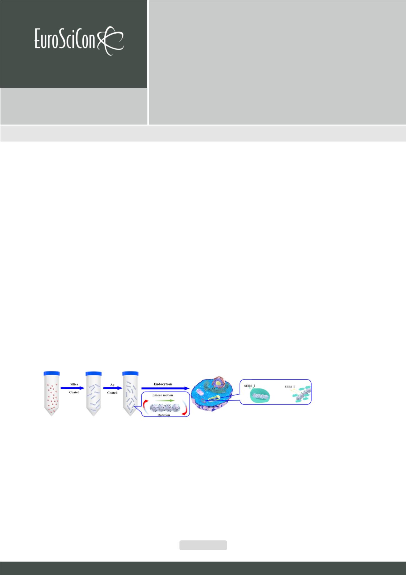
e have fabricated a magnetic SERS probe for biomedical detection
through a handy modified Stöber reaction. The silica reaction is occurred
under magnetic field to align the magnetic particles and string them together
to formmicro rob structure. Its geographical magnetic moment offers a perfect
platform for precise movement and locomotion manipulation under magnetic
field. We can make it go through a micro scaled maze easily to reach targeted
area via magnetic guiding and driving. Not only it could move as we designed
but also it could rotate with demanding angular velocity for some very special
application such as cell tissue depletion experiments. Its surface satellite
doping silver is just adding SERS (Surface Enhanced Raman Spectrum) as an
extra function for its biomedical detection. The detection ability is checked
by crystal violet
in vitro
. The full potential of biomedical sensor in vivo will be
explored in view. Its multi-functional ability makes it an outstanding candidate
for further biomedical application such as micro surgery robot, biomedical
sensor and of course targeted drug delivery mediation
Biography
Huanhuan Feng has completed his PhD from Wageningen
University and Postdoctoral studies in Wageningen University.
He is an Assistant Professor of School of Materials Science
and Engineering, Harbin Institute of Technology (Shenzhen). He
has published more than 20 papers in reputed journals and has
been serving as an Editor of Open Chemistry, De Gruyter.
fenghuanhuan@hit.edu.cnA magnetic SERS probe fabrication and in cell
Huanhuan Feng
1
, Yuhuan Liu
1
, Tingting Zheng
2, 3
and Xing Ma
1
1
Shenzhen Graduate School of Harbin Institute of Technology (HITSZ), China
2
PKU-HKUST, China
3
Peking University Shenzhen Hospital, China
Huanhuan Feng et al., Nano Res Appl Volume:4
DOI: 10.21767/2471-9838-C6-024
Figure 1:
Schema of SERS probe fabrication and experiments in cell