Keywords
Diabetes Mellitus, Experimental; Image Cytometry; Muscle, Skeletal; Muscles; Quadriceps Muscle; Rats
Abbreviations
FBS: fasting blood sugar; STZ: streptozotocin
INTRODUCTION
The concept of insulin action on the skeletal muscles is of great significance in glucose homeostasis, especially in the treatment of type II diabetes. Normally, about 75% of the total body glucose uptake stimulated by insulin is mediated by the skeletal muscles [1, 2]. Insulin action on skeletal muscle fibers takes place via specific membrane receptors coupled to tyrosin kinase activity which eventually leads to glucose uptake by activated GLUT 4 transporters [2]. A large number of reports on the physiology of skeletal muscle fibers in human type I diabetes and in experimentally induced streptozotocin- (STZ) diabetes have revealed a synaptic delay at the motor end plate with a decrease in the contractility of the fibers [3, 4, 5, 6]. Histochemical and morphologic studies with light and electron microscopes have illustrated degeneration and necrosis of the myofibers with atrophy of type 2A and 2B myofibers in the hindlimb muscles of STZdiabetic mice and rats [7, 8], and myofibril derangement and mitochondrial swelling in the hindlimb muscles of spontaneous diabetic WBN/Kob rats [9]. Recent studies on human type II diabetes have illustrated atrophy in the distal muscles of the lower limbs [10]. Several cases of acute skeletal muscular infarction in type II diabetics which are attributed to microangiopathic and coagulopathic causes induced by prolonged lack of insulin have been reported [11, 12, 13]. Reviewing the literature, it seems that few quantitative analytic studies on diabetic skeletal muscles have been conducted. Therefore, the aim of this study was to comprehensively analyze the morphometric changes in the number and size of myofibers and their myonuclei in the extensor digitorum longus and rectus femoris muscles of STZinduced diabetic rats. A comparison in the morphometric data between forelimb and hindlimb muscles was also carried out in order to point out any regional differences in the effect of insulin depletion.
METHODS
Twelve adult male albino Fischer-344 rats (about 300 g in body weight) were used in the study. A group of 6 rats received citrate buffer as a drug vehicle and were maintained on normal food pellets and water while another group of 6 rats were made diabetic by a single intravenous injection of STZ (75 mg/kg body weight) in 0.05 M citrate buffer (pH 4.5) via the tail vein. The STZ-injected rats were kept in separate cages and maintained on the same food pellets. The diabetic status in the STZinjected animals was confirmed from the elevated blood glucose levels and markedly positive glucosuria. Body weight, blood glucose and glucosuria in the normal and diabetic rats were regularly recorded until sacrifice. Four weeks after the onset of the experiment, normal and STZ-diabetic rats were sacrificed under nembutal anesthesia after recording their body weight, fasting blood sugar level and glucosuria. Blood samples for serum insulin assay were taken, the fore and hind limbs of each rat were dissected and samples from the central part of the extensor digitorum longus and rectus femoris muscles were cut, cleaned of fibrous tissue and fixed in modified Bouin’s fluid (picric acid:formaldehyde 3:1) for 24 h at room temperature with continuous shaking. The tissue samples dehydrated in graded ethanol series were embedded in paraffin with proper orientation for cross and longitudinal sectioning.
For the morphometric analysis, an unbiased sampling procedure was applied. Four tissue blocks for cross and longitudinal sectioning per animal were randomly selected, and serial sections of 4 μm thickness were cut and mounted on egg albumin-coated slides. All sections were processed for routine histological examination, stained with hematoxylin-eosin, and examined with a light microscope (Olympus, Tokyo, Japan). One cross and one longitudinal section per block were selected, and three microscopical fields per section were randomly selected for measuring selected parameters. The mean diameter of the muscle fibers and the length of the myonuclei per field were measured from longitudinal sections while the mean number of the muscle fibers, the diameter of the muscle fibers, the number of myonuclei and the diameter of the myonuclei per field were recorded from the cross sections. The diameters of the muscle fibers and myonuclei from the cross sections were calculated from their major and minor profile semi-axes according to the formula of Abercrombie [14]. All quantitative measurements were carried out with a100x objective lens using a calibrated length reticule.
The serum insulin level from each rat was measured by means of the microparticle enzyme immunoassay (MEIA) method using AxSYM insulin assay apparatus (Abbott Laboratories, Abbott Park, IL, USA).
ETHICS
The rats were bred at Jordan University Animal House following the principles of laboratory animal care according to the criteria outlined in the "Guide for the Care and Use of Laboratory Animals (1996)" prepared by the National Academy of Sciences.
STATISTICS
Data from each animal were estimated separately (n=4 blocks), and the mean data were pooled for the entire group of normal and STZ-diabetic rats (n=6 animals in each group). The data are presented as means with standard error of the mean (SEM), and they were analyzed with the Student’s t-test. Data were analyzed by means of the SPSS Version 12 for Windows. Two-tailed P values less than 0.05 were considered statistically significant.
RESULTS
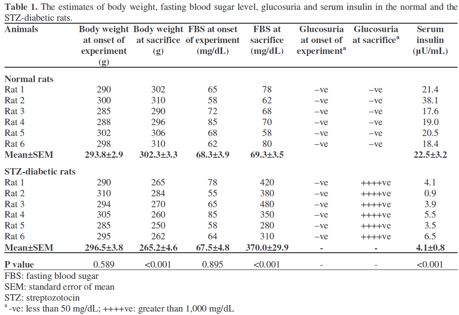
The STZ-diabetic rats showed a significant decrease in their body weight with marked hyperglycemia and positive glucosuria. The serum insulin level was remarkably reduced in streptozotozin-diabetic rats. Table 1 illustrates the values of body weight, fasting blood sugar, glucosuria and serum insulin levels for each rat in the control and the diabetic groups.
Histological examination of hematoxylineosin stained sections from the extensor digitorum longus and rectus femoris of normal and diabetic rats showed adequately preserved myofibers with clear striation and peripheral myonuclei (Figures 1a-d and 2a-d). Areas of inflammation infiltrating the fibers of both muscles were seen in the STZ-diabetic rats (Figure 3a). These zones showed an increase in the number of blood capillaries with many inflammatory cells invading the intercellular spaces. Occasionally, small areas of necrosis and lyses were seen among normal looking fibers (Figure 3b).
Figure 1. H&E stained sections of the extensor
digitorum longus muscle. a. Cross section of a normal
rat. b. Cross section of a STZ-diabetic rat. c. Longitudinal section of a normal rat. d. Longitudinal
section of STZ-diabetic rat. (Magnification x400).
Figure 2. H&E stained sections of the rectus femoris
muscle. a. Cross section of a normal rat. b. Cross
section of a STZ-diabetic rat. c. Longitudinal section of
a normal rat. d. Longitudinal section of STZ-diabetic
rat. (Magnification x400).
Figure 3. a. H&E stained section of the extensor
digitorum longus muscle in a STZ-diabetic rat showing
an area of inflammatory cell infiltration among the
normal myofibers. b. H&E stained section of the rectus
femoris muscle in an STZ-diabetic rat showing
necrosis of the myofibers. (Magnification x400).
Table 2 illustrates the morphometric data of parameters measured from all fields analyzed in the longitudinal and cross sections of the extensor digitorum longus in one of the normal rats.

Table 3 illustrates the mean data obtained from the longitudinal and cross sections of the extensor digitorum longus muscle in the normal and STZ-diabetic rats. The diameters of the muscle fibers estimated from the longitudinal sections was 36% lower in diabetic rats (20.6±1.3 μm versus 32.4±0.2 μm, P<0.001). Similarly, the diameter of the muscle fibers estimated from the cross sections was 31% lower in STZ-diabetic rats (29.0±0.2 μm versus 42.0±1.1 μm, P<0.001) with a 13% increase in their numerical density (14.6±0.5 versus 12.7±0.3, P=0.005). The lengths and diameters of the myonuclei in the diabetic rats were slightly reduced in both muscle fibers which amounted to 4% (14.4±0.2 μm versus 14.6±0.1 μm, P=0.384) and 6% (4.3±0.2 μm versus 4.4±0.03 μm, P=0.783), respectively. A 10% decrease in the numerical density of the myonuclei from the cross sections of the diabetic rats was also recorded (16.5±0.7 versus 18.4±0.7, P=0.094).

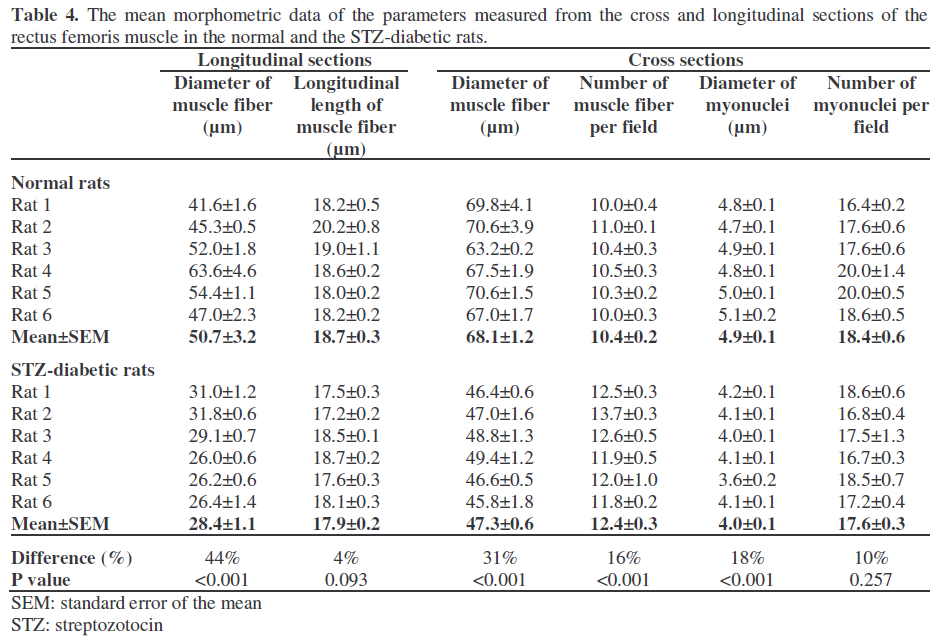
Table 4 records the data obtained from the longitudinal and the cross sections of the rectus femoris muscle in both normal and STZ-diabetic rats. The mean diameter of the fibers in the longitudinal sections was 44% lower in diabetic rats (28.4±1.1 μm versus 50.7±3.2 μm, P<0.001). In the cross sections, the reduction in the diameter of the muscle fibers of the diabetic rats was 31% (47.3±0.6 μm versus 68.1±1.2 μm, P<0.001), and their numerical density increased by 16% (12.4±0.3 versus 10.4±0.2, P<0.001) as compared to the data of the normal rats. The analysis of the myonuclei in the diabetic rats revealed a 4% reduction in their length (17.9±0.2 μm versus 18.7±0.3 μm, P=0.093) and an 18% reduction in their diameter (4.0±0.1 μm versus 4.9±0.1 μm, P<0.001). The numerical density of the myonuclei from the cross sections was 10% lower in the diabetic rectus femoris (17.6±0.03 versus 18.4±0.54, P=0.257).
DISCUSSION
Muscular atrophy and necrosis are well known complications of longstanding human diabetes, and commonly affect the lower limb muscles [11, 12]. This has been attributed to microangiopathies and abnormal coagulation in the skeletal muscle blood vessels caused by persistent hyperglycemia. Most of these cases have been diagnosed from clinical features and radiological imaging procedures, such as MRI, computed tomography, sonography and gallium scintigraphy [12]. Although, 95% of the cases of human diabetic muscular infarction were confirmed by biopsies, physicians usually prefer noninvasive methods and spare muscle biopsy for atypical and uncertain cases of musclular infarction. Based on this information and from a review of the literature, our study was aimed at exploring the morphological changes in the skeletal muscles of the fore and hind limbs of STZ-diabetic rats using quantitative morphometry under a light microscope. Large, randomly selected samples from the extensor digitorum longus and rectus femoris muscles were analyzed using both longitudinal and cross sections. The size and number of the myofibers and the myonuclei were quantified in both muscles of the normal and the STZ-diabetic rats. The results indicate a reduction in the diameter of the fibers with a corresponding increase in their numerical density per unit of reference area. Similarly a slight decrease in myonuclei length and diameter was also recorded in STZ-diabetic rats. These findings were more obvious in the rectus femoris muscle.
Very few quantitative morphometric studies on the skeletal muscles of STZ-diabetic animals have been published. Hegarty and Rosholt [15], Fahim et al. [5], Klueber and Feczko [7], and Ozaki et al. [9], in their studies on different muscles of STZ-diabetic mice and rats, have reported a decrease in the number and diameters of the muscle fibers. Studies on human type II diabetes have illustrated impaired skeletal muscle strength with a decreased muscle area, and infarctions [4, 11]. Studies on different muscles from STZ-diabetic rats have shown a decrease in protein synthesis and an increase in ribosomal degradation associated with changes in the circulating and muscle amino acids [16, 17]. Chaudhary et al. [18], from their study on the fore and hindlimb muscles of STZ-diabetic rats, have demonstrated a decrease in the weight and protein content of fast conducting muscles. They concluded that diabetic atrophic changes in skeletal muscles vary according to fiber composition and function. Similarly, Kawaguchi et al. [19], from their work on skeletal and cardiac muscles in human diabetes and in STZ-diabetic rats, observed small myocytes due to decreased Factin production.
In the course of this study, patches of inflammatory reaction were occasionally seen infiltrating the myofibers of the STZ-diabetic rats. Torres et al. [20], in their study on noncomplicated type II diabetic patients, recorded an increase in nitric oxide production and elevated levels of CD163, CD154 and TNFalpha in samples of quadriceps femoris muscles. This was associated with macrophage infiltration and concluded that an inflammatory process occurs in the skeletal muscles of diabetic patients. Similar inflammatory changes in the skeletal muscles have also been reported in STZ-diabetic rats [21].
The process of skeletal muscle atrophy caused by insulin depletion is rather complicated and seems to involve several overlapping mechanisms including metabolic derangement, blood vessel changes, motor end plate degeneration and impairment of myocyte protein synthesis. In conclusion, our study clearly illustrates a decrease in the size of the myofibers in the fore and hind limb muscles of STZ-diabetic rats, there being a more prominent effect in the hindlimb muscles. Further studies on diabetic muscular atrophy and peripheral neuropathy are required to elucidate and understand the role of insulin as a causative agent.
References
- Bjornholm M, Zierath JR. Insulin signal transduction in human skeletal muscles: identifying the defects in Type II diabetes. BiochemSoc Trans 2005; 33:354-7. [PMID 15787605]
- Idris I, Gray S, Donnelly R. Insulin action in skeletal muscles: isozyme-specific effects of protein kinase C. Ann N Y AcadSci 2002; 967:176-82. [PMID 12079846]
- Andersen H, Schmitz O, Nielsen. Decreased isometric muscle strength after acute hyperglycaemia in Type 1 diabetic patients. Diabet Med 2005; 22:1401- 7. [PMID 16176203]
- Cederholm T, Sylven C, Esbjornsson-Liljedahl M, Jansson E. Insulin treatment increases skeletal muscle fiber area in patients with diabetes mellitus type 2. ClinPhysiol 2000; 20:354-9. [PMID 10971546]
- Fahim MA, el-Sabban F, Davidson N. Muscle contractility decrement and correlated morphology during the pathogenesis of streptozotocin-diabetic mice. Anat Rec 1998; 251:240-4. [PMID 9624455]
- Medina-Sanchez M, Rodriguez-Sanchez C, Vega- Alvarez JA, Menedez-Pelaez A, Perez-Casas A. Proximal skeletal muscle alterations in streptozotocindiabetic rats: a histochemical and morphometric analysis. Am J Anat 1991; 191:48-56. [PMID 1829578]
- Klueber KM, Feczko JD. Ultrastructural, histochemical, and morphometric analysis of skeletal muscles in a murine model of type I diabetes. Anat Rec 1994; 239:18-34. [PMID 8037375]
- Chao TT, Ianuzzo CD, Armstrong RB, Albright JT, Anapolle SE. Ultrastructural alterations in skeletal muscle fibers of streptozotocin-diabetic rats. Cell Tissue Res 1976; 168:239-46. [PMID 131648]
- Ozaki k, Matsuura T, Narama I. Histochemical and morphometrical analysis of skeletal muscle in spontaneous diabetic WBN/Kob rat. ActaNeuropathol (Berl) 2001; 102:364-70. [PMID 11585251]
- Greenman RL, Khaodhiar L, Lima C, Dinh T, Giurini JM, Veves A. Foot small muscle atrophy present before the detection of clinical neuropathy. Diabetes Care 2005; 28:1425-30. [PMID 15920063]
- Ran X, Wang C, Wang H, Zhao T, Tong N, Song B, et al. Muscle infarction involving muscles of abdominal and thoracic walls in diabetes. Diabet Med 2005; 22:1757-60. [PMID 16401324]
- Trujillo-Santos AJ. Diabetic muscle infarction: an underdiagnosed complication of long-standing diabetes. Diabetes Care 2003; 26:211-5. [PMID 12502683]
- Reyes-Balaguer J, Solaz-Moreno E, Morata-Aldea C, Elorza-Montesinos P. Spontaneous diabetic myonecrosis. Diabetes Care 2005; 28:980-1. [PMID 15793211]
- Abercrombie M. Estimation of nuclear population from microtome sections. Anat Rec 1946; 94:239-47.
- Hegarty PV, Rosholt MN. Effects of streptozotocin-induced diabetes on the number and diameter of fibres in different skeletal muscles of the rat. J Anat 1981; 133:205-11. [PMID 7333950]
- Rodriguez T, Alvarez B, Busquets S, Carbo N, Lopez-Soriano FJ, Argiles JM. The increased skeletal muscle protein turnover of the streptozotocin-diabetic rat is associated with high concentrations of branchedchain amino acids. BiochemMol Med 1997; 61:87-94. [PMID 9232202]
- Ashford AJ, Pain VM. Effect of diabetes on the rates of synthesis and degradation of ribosomes in rat muscles and liver in vivo. J BiolChem 1986; 261:4059-65. [PMID 2419338]
- Chaudhury SK, Mandal MB, Deshpande SB, Saxena ID. Effect of streptozotocin-induced diabetes on growth and proteolytic activity of different muscles in rats. Indian J ExpBiol 1994; 32:877-80. [PMID 7896320]
- Kawaguchi M, Asakura T, Saito F, Nemoto O, Maehara K, Miyake K, et al. Changes in diameter size and F-actin expression in the myocytes of patients with diabetes and streptozotocin-induced diabetes model rats. J Cardiol 1999; 34:333-9. [PMID 10642930]
- Torres SH, De sanctis JB, de L Briceno M, Hernandez N, Finol HJ. Inflammation and nitric oxide production in skeletal muscle of type 2 diabetic patients. J Endocrinol 2004; 181:419-27. [PMID 15171690]
- Perreault M, Dombrowski L, Marette A. Mechanism of impaired nitric oxide synthase activity in skeletal muscle of streptozotocin-induced diabetic rats. Diabetologia 2000; 43:427-37. [PMID 10819235]


