Keywords
Fascioliasis; Sheep; Cattle; Goat; Liver; Slaughter house
Introduction
Definitely, fascioliasis is among the most widely distributed zoonotic diseases, and it is a serious economic disease of cattle and sheep with a worldwide distribution. However, affecting no less than 300 million cattle and 250 million sheep throughout the world. Also, it is a common parasitic disease in ruminants, especially in cattle, sheep, buffaloes, swine and goats. It may also affect humans [1,2]. Fasciola spp. has two main types, they are: F. hepatica and F. gigantica. Differentiation of these types can be through morphological features like shape and body length [3]. F. hepatica has a complex developmental life cycle [4]. This huge economic impact forming direct losses might be an underestimate considering indirect costs of treatment, or loss of animal workforce in less industrialized countries. Furthermore, fascioliasis is emerging as a relevant issue in human health, affecting roughly 2.6 million people worldwide. For this reason, it has been considered as a re-emerging neglected disease by the WHO [2].
Stage of the bisexual adult is established in the host bile ducts and reproduced sexually, discharging thousand eggs daily. These eggs are passing within bile ducts into intestines, and then spreading in the stream of fecal [2,5]. Eggs which reach freshwater embryonate over two weeks, producing a freeswimming miracidium. This miracidium is looking for and infecting a Lymnaeidae family snail. The parasite inside the snail advances through stages of sporocyst, redia, and daughter redia by non-sexual reproduction and growth, releasing thousand cercaria [5]. The free-living, aquatic cercaria encysts as the metacercarial stage on solid substrates, including vegetation at the margins of watercourse. When an appropriate host consumes an infected vegetation (e.g., uncooked watercress), the metacercarial stage excysts in the duodenum, transverses the small wall of intestine, transfers through the peritoneal cavity, and penetrates the Glisson's capsule of liver [1].
The liver is damaged by the incidental fluke migration through the parenchyma of liver into the biliary ducts. This would stimulate reactions related to the severe infection phase. Systemic diseases accompany this phase, comprising abdominal pain, nausea and fever. Diseases like inflammation, anaemia, fibrosis, biliary stasis and cholangitis may inaugurate when the adult is established in the bile ducts. During this prolonged phase, mature worms can live numerous years in the intervention obscurity [6,7]. In spite of its vigorous and extensive action against other human parasitic flatworms, no effect is found for the anthelmintic drug praziquantel on F. hepatica [8]. The ecological changes that occurs in vast regions middle and northwest of Iraq is one outcome of military conflicts that were encountered between the year 2014 and 2015 when Several dams and water systems were destroyed accompanied by floods followed by a period of drought [9]. Kirkuk district was selected as an example of such environmental changes to identify the impacts of these changes on the prevalence of fascioliasis in local ruminants [10,11]. This paper mainly focused on the liver flukes, F. hepatica which infects ruminants and other mammalian extensively. It aimed at showing the impact of F. hepatica on ruminants represented by causing major diseases to cattle, which in turn produces considerable losses in economy.
Materials and Methods
Study area
This study is a reviewing survey, carried out from 1 September 2017 to 30 April 2018. It investigated records of daily condemnation for sheep, cattle, goats and buffaloes in the biggest slaughterhouse (Aleasria) in Kirkuk (East province, north of Iraq). Kirkuk lies in the latitude 35.478565, and longitude 44.401932, with coordinates of GPS as 35° 28' 42.8340 N and 44° 24' 6.9552 E. Economy of the city is based on agriculture, that most people around Kirkuk are farmers. The city is famous for being the Food Basket of the Nation (Iraq) due to its various rich agricultural productions, including: corn, wheat, barley, tomatoes and sunflower etc. Above 5% of population are involved in arable areas. Scientific committee at Veterinary Medicine College/ Kirkuk University (Iraq) has approved this study. Cattle were raised mostly under seasonal confinement, tethering and semi-extensive system.
Sample collection and animal selection
The study was conducted on different animals (cattle, sheep, buffaloes and goat) in Aleasria slaughterhouse, situated at the south east part of Kirkuk. This slaughterhouse contains two main halls: one is for sheep accommodating up to (40488) slaughtered sheep; while the other one is for cattle accommodating up to (11707) slaughtered cattle; besides, it has two rooms for slaughtered goats and buffaloes (3754 and 187), respectively. Additionally, there is a room for authorities of veterinary and another room for changing wears. All animals were from north of Iraq with local origin and slaughtered according to the Islamic way. The abattoir under study sourced its sheep and cattle mostly from neighbouring cities, such as Tikrit, Mosul, Sulaymaniyah, adding to Northern cities in the country. Whereas the source of most goats was from Kirkuk, buffaloes were from south of Iraq. Meat of the four animals under study was mostly inspected by professionals in the field of animal health. Veterinarians were called when having difficulties. Concerning the observation system, inspectors of veterinary meat examined each slaughtered animal during their routine responsibilities. Reasons for disapproval of tissues including fascioliasis were noted daily on prepared data sheets. The F. hepatica was distinguished by faecal samples were taken and stored without fixatives at 4? until they were analysed by coproscopy. A standard sedimentation technique was performed using 10 g of faeces for the detection of F. hepatica eggs [12]. For many of these faecal samples the coproscopy examination was repeated two times in an attempt to improve the sensitivity of the test. The current experimental setup does not have a close resemblance to other information. Finally, demographic data were recorded routinely.
Statistical analysis
A monthly basis was employed to organize the incidence. A software called Sigma Plot (version 12) was used to analyse the data. Special calculation forms were used to record the data. Microsoft® Excel (2013) was used to conduct the initial analysis. There are several possible explanations for result variables of fascioliasis detected during routine postmortem inspection. Calculation of fascioliasis prevalence was performed; then number of animals infected with F. hepatica was stated in a ratio of the total number of formerly chosen animals. Assessment of strong economic consequence of this problem was based on the total number of livers condemned with fascioliasis and that of animals slaughtered in the period of study, monthly calculated, using present data obtained from the abattoir. Cattle were raised mostly under seasonal confinement, tethering and semiextensive system.
Results
This study aimed at investigating fascioliasis prevalence in Aleasria slaughterhouse in Kirkuk. In this slaughterhouse, there were 40488 slaughtered sheep, 3754 goats, 11077 cattle, and then 187 buffaloes (Figure 1). The total number of affected animals with fascioliasis were recorded highest in November: 69 animals, followed by January: 59 animals, October: 44 animals, December: 38 animals, February: 26 animals, September: 21 animals and April: 20 animals; however, the lowest record was in March: 18 animals (Figure 2).
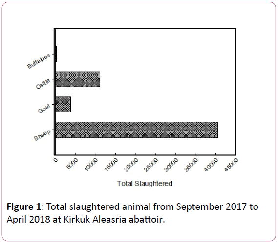
Figure 1: Total slaughtered animal from September 2017 to April 2018 at Kirkuk Aleasria abattoir.
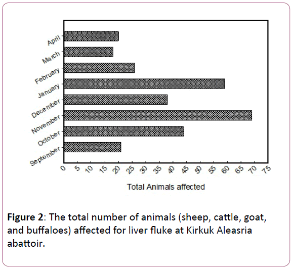
Figure 2: The total number of animals (sheep, cattle, goat, and buffaloes) affected for liver fluke at Kirkuk Aleasria abattoir.
The range of total slaughtered sheep was organized monthly, as follows: April 6457 > September 6145 > November 5620 > January 4794 > February 4678 > March 4672 > October 4300 > December 3822 (Figure 3A). While goats slaughtered was as follows: September 731 > January 520 > November 478 > April 477 > February 447 > March 399 > October 380 > December 324 (Figure 3B). Cattle slaughtered were as follows: September 1657 > April 1624 > November 1597 > January 1357 > February 1299 > March 1282 > October 1161 > December 1100 (Figure 3C). For buffaloes, it was as follows April 32 > November 31 > March 29 > October 28 > February 23 > September 8; whereas similarity was recorded for January and December 18 (Figure 3D).
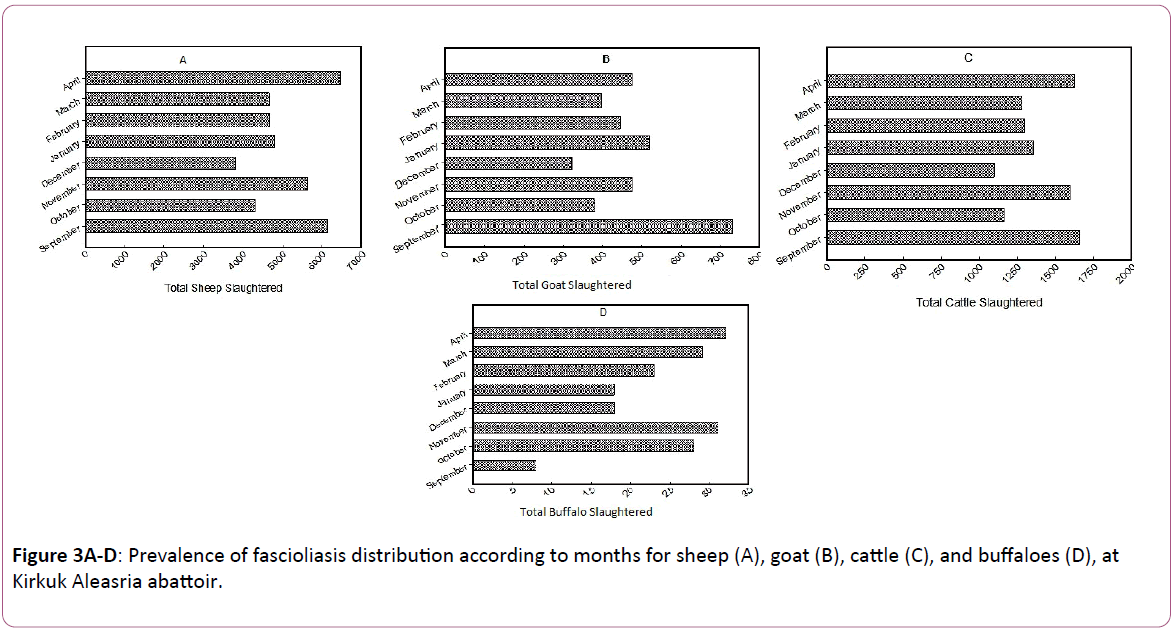
Figure 3 A-D: Prevalence of fascioliasis distribution according to months for sheep (A), goat (B), cattle (C), and buffaloes (D), at Kirkuk Aleasria abattoir.
A total of 114 out of 40488 sheep were observed giving a prevalence of 0.28%; 24 out of 3754 goats with prevalence of 0.63%; 150 out of 11077 cattle with prevalence of 1.35%, and 9 out of 187 buffaloes with prevalence 4.8% in Aleasria slaughterhouse in Kirkuk (Table 1). A total of prevalence in sheep was highest in March 72% and lowest in September 9.5% (Figure 4A). Similarly, prevalence in goat was highest in March 11% and there were no samples recorded in September (Figure 4B). Prevalence in cattle was highest in September 85% and lowest in March 27% (Figure 4C); whereas no prevalence was recorded for buffaloes in January, March, and April (Figure 4D).
| Animal species |
Overall total slaughtered |
Total infection liver fluke |
Prevalence (%) |
P (t) values |
| Sheep |
40488 |
114 |
0.28 |
P=0.451; t=1.168 |
| Goats |
3754 |
24 |
0.63 |
P=0.331; t=0.601 |
| Cattle |
11077 |
150 |
1.35 |
P=0.456; t=0.975 |
| Buffaloes |
187 |
9 |
4.8 |
P=0.278; t=0.412 |
The P values are represent significant differences between months and animals. t values was always positive between affected and non-affected animals
Table 1: Incidence of fascioliasis among slaughtered ruminant in Kirkuk Aleasria abattoir.
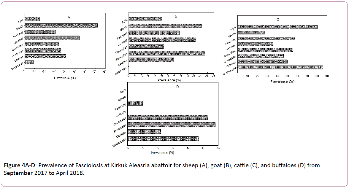
Figure 4 A-D: Prevalence of Fasciolosis at Kirkuk Aleasria abattoir for sheep (A), goat (B), cattle (C), and buffaloes (D) from September 2017 to April 2018.
Polar scatter showed highly R levels for cattle and sheep 114 and 90, respectively; whereas lower R levels for goat and buffaloes were 19 and 8, respectively (Figure 5). No linear regression was observed between positive and negative animals (R2=0.353); also, no significant differences between positive and negative animals (P=0.405) (Figure 6). The macroscopic pathological changes of infected livers of cattle (Figure 7) were characterized by large size. These changes were amplified in a clear and well-marked changes macroscopic sore colour, especially in the case of large size of liver fluke. The colour of liver was dark red with the appearance of a white ring around the worms because of the inflammatory processes that occurred in the liver tissues as well as increased fibrous tissue between lobes of the liver with the presence of necrotic patches among worms in some areas (Figure 7).
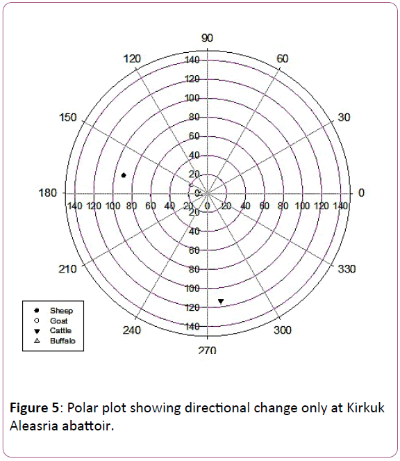
Figure 5: Polar plot showing directional change only at Kirkuk Aleasria abattoir.
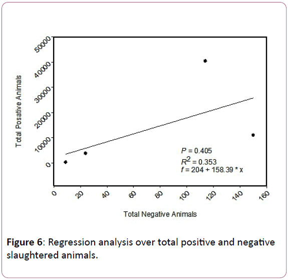
Figure 6: Regression analysis over total positive and negative slaughtered animals.
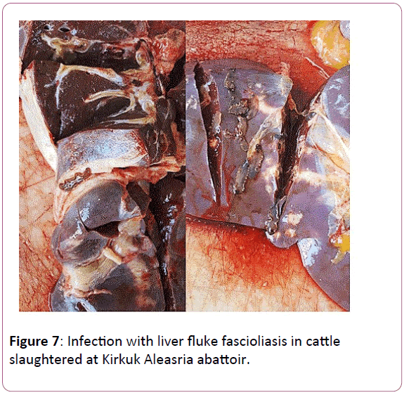
Figure 7: Infection with liver fluke fascioliasis in cattle slaughtered at Kirkuk Aleasria abattoir.
Discussion
A considerable economic loss in cattle is caused by Hepatic fluke infections because of organs condemnation and decrease in the production of meat and milk. So, it is recommended that the effective assessment service of meat must serve as a significant observer of diseases of animal, especially the chronic condition which is not apparent to the owner or the veterinarian [13]. Fascioliasis, as a result of liver fluke species of the genus Fasciola, has usually been nicely diagnosed because of its excessive veterinary impact, but some of the maximum illnesses were omitted for decades with reference to human contamination. However, the increasing significance of human fascioliasis international has re-released interest in fascioliasis. From the Nineteen Nineties, many new ideas had been developed regarding human fascioliasis and these have provided a new model for the human sickness. This is very special to a simple extrapolation from fascioliasis in farm animals [14].
Overall, number of ruminants infected with F. hepatica in Kirkuk (as illustrated in Table 1) was less than Basra province (southern of Iraq) [15] in all animal species. As compared with Iran and Pakistan (Kashmir region), a higher rate was found in a neighboring country in all animal species [8,16]. In Khuzestan Province (Iran), the Fasciola infection in livestock was higher than that found in Saudi Arabia (for all animal species), and in Turkey (only for cattle) [17,18].
The infection rate in November, December, and January was highest (Figures 2-4) compared to other months. This can be attributed to the higher humidity that is recorded in winter which favours the parasites growth [19]. The distribution of infection was low. The lower rate of the infection was because the intermediate hosts (snails) rate in Kirkuk was low. Evidently, the distribution of snails was only in Al-Hawija district, Kirkuk [20,21]. Previous studies showed that infection rate was lower in calves than in cattle. In a study conducted in Babel by [15], the F. gigantic rate is increased according to animal’s advanced age. This study found that the infection rate in cattle was 3.4% for smaller than 2 years and 59% for bigger than 4 years; whereas in buffaloes, only two cases were recorded from fifty examined animals.
Concerning comparison of the current study findings with other studies, conducted a study in Basra and found that the F. gigantic infection rate was higher in summer, followed by spring. Likewise, [15] carried out a study in Babel and found that this infection rate was higher during summer season and lower during winter season. To sum up, this study generally found that the rate of infection was low due to reducing the infected liver of cattle, goat, sheep and buffaloes. This study has helped illustrating the importance of recording the rates of useful meat in monitoring the potentiality important parasitic diseases.
Conclusions
This study found low prevalence of fascioliasis and a great reduction of liver censures in cattle, sheep, goats and buffaloes. This inspection has helped illustrating the utility of records of meat inspection in observing situations of disease and establishing potential extended term trends. The study found that winter is crucial for animals as a result of its highest rates of infection, followed by monsoon and summer seasons. This is as a result of the highest humidity recorded in winter as compared to former years. The highest humidity is considered as the ideal conditions of favourable survive for parasite owing to the prolonged rainfall during winter; therefore, the intermediate host (snail) will increase in Winter in Iraq. Also, this study provides an initial standard data for monitoring these potentially important parasitic diseases in Kirkuk in future.
Acknowledgments
The current study was funded by College of Veterinary Medicine, Kirkuk University, Kirkuk, Iraq. Authors introduce their thanks to the staff of Public Health Department, Veterinary Medicine College for their help. The authors also thankful for the staff of meat inspection and slaughterhouse, especially Dr. Fadhil Abass, in relation to gathering the required data for the current investigation, their effort is highly appreciated.
References
- Burden D, Bland A, Hammet N, Hughes D (1983) Fasciola hepatica: migration of newly excysted juveniles in resistant rats. Exp Parasitol 56:277-288.
- Fürst T, Duthaler U, Sripa B, Utzinger J, Keiser J (2012) Trematode infections: liver and lung flukes. Infect Dis Clin 26:399-419.
- Keiser J & Utzinger J (2009) Food-borne trematodiases. Clin Microbiol Rev 22:466-483.
- Ashrafi K, Valero M, Panova M, Periago M, Massoud J et al. (2006) Phenotypic analysis of adults of Fasciola hepatica, Fasciola gigantica and intermediate forms from the endemic region of Gilan, Iran. Int J Parasitol 55:249-260.
- Rondelaud D, Belfaiza, M, Vignoles P, Moncef M, Dreyfuss, G. (2009) Redial generations of Fasciola hepatica: a review. J Helminthol 83:245-254.
- Marcos L, Terashima A, Leguia G, Canales M, Espinoza J (2007) La infección por Fasciola hepatica en el Perú: una enfermedad emergente. Revista de Gastroenterología del Perú, 27:389-396.
- Lopez M, White Jr A.C, Cabada M.M (2012) Burden of Fasciola hepatica infection among children from Paucartambo in Cusco, Peru. The Am J Trop Med Hyg 86:481-485.
- Chai J.Y (2013) Praziquantel treatment in trematode and cestode infections: an update. J Infect Chemother 45:32-43.
- Shamout M.N & Lahn G. (2015) The Euphrates in crisis: channels of cooperation for a threatened river. Chatham House for the Royal Institute of International Affairs.
- Kadir M & Rasheed S. (2008) Prevalence of some parasitic helminths among slaughtered ruminants in Kirkuk slaughter house, Kirkuk, Iraq. Iraqi J. Vet. Sci, 22:81-85.
- Abed F (2012) A Pathological study of lesions in the liver of sheep in abattoir of Kirkuk province. Europ. J. Appl. Sci, 4:140-145.
- Rapsch C, Schweizer G, Grimm F, Kohler L, Bauer C, et al. (2006) Estimating the true prevalence of Fasciola hepatica in cattle slaughtered in Switzerland in the absence of an absolute diagnostic test. Int J Parasitol 36:1153-1158.
- Blamire R, Goodhand R, Taylor K (1980) A review of some animal diseases encountered at meat inspections in England and Wales, 1969 to 1978. The Veterinary record, 106:195-199.
- Mas-Coma S, Valero M.A, Bargues M.D (2009) Fasciola, lymnaeids and human fascioliasis, with a global overview on disease transmission, epidemiology, evolutionary genetics, molecular epidemiology and control. Adv Parasitol 69:41-146.
- Mathur P, Al-Fathy F, Karim M (1974) Observation on the incidence of larval cestodes of veterinary and public health importance in northern Iraq. Working paper, UNDP/FAO, Development of Livestock Production in northern Iraq, IRQ 71/542. Google Scholar
- Ahmadi N.A, Meshkehkar M (2010) Prevalence and long term trend of liver fluke infections in sheep, goats and cattle slaughtered in Khuzestan, southwestern Iran.
- Over H.J, Jansen J, Van Olm P. (1992) Distribution and impact of helminth diseases of livestock in developing countries. Food & Agriculture Org.
- Gargili A, Tüzer, E, Gülenber A, Toparlak M, Efil ? et al. (1999) Prevalence of liver fluke infections in slaughtered animals in Trakya (Thrace), Turkey. Turk. J. Vet. Anim. Sci 23:115-116.
- Pandya S.S, Hasnani J.J, Patel P, Chauhan V.D, Hirani N.D et al. (2015) Study on prevalence of Fasciolosis in buffaloes at Anand and Ahmedabad districts, Gujarat, India. Veterinary world, 8:870.
- Mahdi N, & Al-Baldawi F (1987) Hepatic fascioliasis in the abattoirs of Basrah. Ann Trop Med Parasitol 81:377-379.
- Al-Delemi J (2005) Epidrmiological and immunological study for Fasciola gigantica among cattle in Babylon province. Ph. D. thesis, College of Veterinary Medicine, University of Baghdad, Iraq.