Key words
Labor, Parturition, domestic animal, hormones
Introduction
Labor is characterized by an increase in myometrial activity or, more precisely, a change in the myometrial
contractility pattern from "contractures"(long-lasting, low frequency activity) to contractions" (high intensity, high
frequency activity) [14], resulting in effacement and dilatation of the uterine cervix. In other words, Parturition is the
process of delivery of the fully-grown fetus on the completion of the normal pregnancy period. Parturition is an
interesting biological process in the sense that the uterus that was quiescent during the entire pregnancy starts
contracting and the cervix that was tightly contracted relax sufficiently to allow the passage of the young one to the
world outside the mother’s womb, passing through the birth canal (which Is formed by the uterus, cervix and vagina
within the pelvic bones and their attachments). Pre parturient Care of the mother throughout the gestation and
especially during the last part, the nutrition of the pregnant animals is important. Feeding of animals should be
oriented in such a way that the prepartum or parturient incidence of some of the commonly occurring metabolic
disorders is minimized, a healthy viable progeny is produced and the milk production of the dairy type animals is
optimum. It is beyond the scope of this book to discuss all of these strategies in detail. In dairy cattle, farmers often
feed their pregnant cows with concentrates only during the last few days of pregnancy and often vegetable oil is
added to the concentrates. Although growth of the fetus occurs maximally during the last part of gestation, however,
the value of such oil feeding is not beyond doubt. Recent suggestions for feeding of pregnant dry cows include the
feeding of high-fiber low-energy chopped straw [13,15] and the feeding of anionic salts in combination with
adequate calcium and magnesium [4] and restriction of rumen degradable protein [14]. Extra energy feed is required
for sheep and goats that have been diagnosed to be carrying twins. The feeding of the bitch should be aimed at increasing the energy intake during the last four weeks of pregnancy and 1.0 –1.8% calcium and 0.8-1.6% of
phosphorous should be included in the diet of late pregnant bitches [1]. Vaccination of pregnant animals for the
prevention of some infectious diseases has been mentioned previously, however, these vaccinations depend on
whether or not, the disease is prevalent and the species-specific requirement. Pregnant mares however, need to be
essentially given tetanus antitoxin or tetanus toxoid during gestation and immediately after foaling. Special attention
need to be attached to the hygiene at the time of parturition and as such, animals must be shifted to hygienic
parturition stalls and this would also prevent overcrowding.
Signs of approaching parturition
Some externally visible changes do occur in animals when parturition is approaching. The most important external
changes of approaching parturition are seen in the udder, vulva and pelvic ligaments and to some extent in the
behavior. The symptoms are inconsistent between individual animals, and between consecutive parturitions. The
symptoms therefore, do not permit an accurate prediction as to the exact time of parturition in a certain animal but
are only useful indications as to the approximate time parturition can be expected. Clinicians must therefore refrain
from too positive statements concerning the exact time of parturition.
Physiological phases of myometrial activity
The regulation of uterine activity during pregnancy can be divided into four distinct physiologic phases [5,6]: Phase
0: inhibitors active during pregnancy the uterus is maintained in a state of functional quiescence through the action
of various putative inhibitors including, but not limited to:
- Progesterone
- Prostacyclin (prostaglandin I-2)
- Relaxin
- Parathyroid hormone-related peptide Nitric oxide
- Calcitonin gene-related peptide
- Adrenomedullin
- Vasoactive intestinal peptide.
Phase 1: myometrial activation as term approaches the uterus becomes activated in response to uterotropins, such as
estrogen. This phase is characterized by increased expression of a series of contraction-associated proteins (CAPs)
(including myometrial receptors for prostaglandins and oxytocin), activation of specific ion channels, and an
increase in connexin-43 (a key component of gap junctions). An increase in gap junction formation between
adjacent myometrial cells leads to electrical synchrony within the myometrium and allows for effective coordination
of contractions.
Phase 2: stimulatory phase Following activation, the "primed" uterus can be stimulated to contract by the action of
uterotonic agonists, such as the stimulatory prostaglandins E2 and F2 alpha and oxytocin.
Phase 3: involution Involution of the uterus after delivery occurs during phase 3 and is mediated primarily by
oxytocin.
Closure of the ductus arteriosus.
Closure of the foramen ovale within a few hours ofbirth in foal. Prepartum signals for parturition initiation the
initiation of parturition in most domestic animals continues to be only partially understood. It is fascinating that on
completion of events necessary to render a young one capable of independent life outside the mother’s uterus,
closely coordinated changes occur in the fetus and mother resulting into delivery of the fetus by the act of
parturition. Possibly the initial mechanism for the timing of birth is encoded in the fetal genome and is closely
linked to, and activated when certain prerequisite developmental events have occurred in the fetus [5]. The possible
factors that help in initiation and the act of parturition include physical, biochemical and neuro endocrine (Table 1)
factors.
Table 1: Possible factors responsible for initiation of parturition
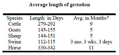
Table 2: Duration of different stages of labor in domestic animals
It is considered that in most species the fetus exerts a control over the length of gestation whereas, the mother can
influence the time of birth within the narrow limits [6]. The fetal pituitary adrenal axis is known to initiate the
prepartum events by which signals to the placenta trigger the maternal hormonal changes which allow normal labor
to proceed at least in the ruminants and to some extent in the pig [15, 16]. The role of fetus and the nature of its
signals to the mother for maternal changes are still unknown in the horse [15] and the dog [3, 4, 8]. The uterus
remains quiescent during pregnancy in most domestic animals by a combined action of luteal and / or placental
progesterone and molecules like relaxin, nitric oxide, prostacyclin and catecholamine’s [8]. This is transformed into
an oscillatory organ with cervical relaxation near parturition. Many biochemical, hormonal and molecular changes
precede parturition. The universality of the fetal glucocorticoid surge (sudden rise in levels) preceding normal labor
at term suggests that it may represent a fundamental signal common to all species [14].
Hormones Involved in the Parturition
Prostaglandins
Prostaglandins are predominantly paracrine/autocrine hormones (i.e., they act locally at their site of production on
contiguous cells). An increase in uterine prostaglandin biosynthesis is a consistent element in the transition into
labor [7], and is probably common to all species [8].
Progesterone
Administration of a progesterone receptor antagonist or removal of the readily induces abortion in early pregnancy
(before 7 weeks of gestation), corpus luteum [suggesting that progesterone is necessary for early pregnancy
maintenance. Administration of exogenous progesterone after early lutectomy prevents abortion, further supporting
the hypothesis that ovarian progesterone production is essential in maintenance of early pregnancy. Placental
progesterone production becomes important between 7 and 9 weeks, and the placenta is the dominant source of
progesterone thereafter. However, the role of progesterone in late pregnancy is not as well defined.
Estrogen
The placenta is the primary source of estrogen biosynthesis during pregnancy. Estrogens do not themselves cause
myometrial contractions, and maternal administration of estradiol to rhesus macaques from 130 days of gestation
has no effect on length of pregnancy [11]. Instead, estrogens act by up regulating myometrial gap junctions [4] and
uterotonic receptors (including L-type calcium channels and oxytocin receptors) [2], thereby enhancing the capacity
of the myometrium to generate contractions.
PGF 2alpha
Enhances myometrial contractions, induces luteolysis and the release/secretion of relaxin.
Oxytocin
Oxytocin is a peptide hormone synthesized in the hypothalamus and released from the posterior pituitary in a
pulsatile fashion. Its biologic half-life is approximately three to four minutes, but appears shorter when higher doses
are infused. Oxytocin is inactivated in the liver and kidney, although during pregnancy it is primarily degraded by
placental oxytocinase. Oxytocin is the most potent endogenous uterotonic agent, and is capable of stimulating
uterine contractions at intravenous infusion rates of 1 to 2 mU/min at term [16,3]. The frequency and amplitude of
oxytocin-induced uterine contractions are identical to those occurring during spontaneous labor.
Glucocorticoids
These hormones have several actions that can also help prepare the uterus for labor. Glucocorticoids act directly to
up regulate prostaglandin production in fetal membranes at term[6,13]. Cortisol appears to stimulate expression of
placental (but not hypothalamic) CRH in vitro [10]. In addition, cortisol enhances amnionic cyclooxygenase to
enhance prostaglandin synthesis and inhibits chorionic prostaglandin dehydrogenase activity, thereby preventing
prostaglandin metabolism [6,1]. Parathyroid hormone-related peptide: Parathyroid hormone-related peptide is a
potent smooth muscle relaxant capable of inhibiting oxytocin-induced contractions in baboons [12]. It is unclear
whether it has a physiologically important role in maintaining uterine quiescence prior to the onset of labor.
Relaxin
Relaxin is a member of the insulin-like growth factor family of proteins. Plasma levels are highest at 8 to 12 weeks
of gestation and thereafter decline to low levels, which persist until term [9]. The primary source of relaxin is
thought to be the corpus luteum.
Parturition
It is very important that during parturition the animal moves smoothly through the three stages of parturition n a
reasonable amount of time to ensure a safe and normal birth. The first stage of parturition is known as the Preparatory Stage. It is during this stage, as is evident from the name that the female prepares to give birth. Some
signs that parturition is near can be observed during this stage. In addition, there may be mucus discharge from the
vulva, decreased body temperature, filling of mammary glands with milk, and mild straining. Behavioral signs
include nesting behavior and possibly stealing other newborns in the herd. The second stage of parturition is the
Expulsion Stage. It is at this point that the walls of the uterus begin to contract more frequently and with increased
force, thus pushing the fetus into the birth canal. When the contractions become strong enough, the fetus is actually
forced out of the female’s body, hence the name the Expulsion Stage. Once an animal has entered this stage,
delivery should occur fairly soon; otherwise, there may be difficulties preventing normal delivery. Again, the
standard amount of time allowed for a normal birth can differ between species. The final stage of parturition is the
Cleaning Stage. It is at this point in the process that the afterbirth, or the placenta, is expelled from the body. In
order for the animal to make a normal, healthy recovery from parturition, the afterbirth must be expelled. If the fetal
membranes and fluid remain in the animal, they can become infected and lead to serious illness and possible death
of the mother.
The Role of the Fetus on beginning
Considerable evidence suggests that in most viviparous animals the fetus controls the timing of onset of labor [15-12, Figure 1]. During the Hippocratic period, the fetus was thought to be positioned head down at term so it could kick
its legs up against the fundus of the uterus, thereby propelling itself through the birth canal. While we have moved
away from this simple and mechanical view of labor, the factors responsible for the initiation and maintenance of
labor at term are not well defined. Initial investigations focused on endocrine events, such as changes in the profile
of circulating hormone levels in the maternal and fetal circulations. Subsequent studies have concentrated on the
dynamic biochemical dialogue between the fetus and mother (paracrine/autocrine events) in an attempt to
understand the molecular mechanisms that regulate such interactions. The genetic regulations of the molecular
events that occur during parturition are also being investigated [5].
Figure 1: The role of fetus on beginning of labor
Pathways to parturition: this section was indicated in Figure 2.
Figure 2: Pathway to parturition
Interaction between: Prostaglandins, Cytokines and Oxytocin
Observations suggest that prostaglandins are positive regulators of oxytocin receptor expression, that the uterotonic
action of prostaglandins depends partly on oxytocin action, and that prostaglandins, but not oxytocin, are directly
involved in the initiation of labor and the role of cytokines is in direct relation on performance of PG. The maximum
number of studies on parturition processes has been conducted on sheep. Progesterone production in the pregnant
sheep is derived from the corpus luteum during the first 50 days of pregnancy [5] but there is a gradual decline in
ovarian progesterone secretion thereafter [4,6]. Thus ovariectomy after day 50 does not cause abortion because
placental progesterone is adequate to maintain pregnancy [6]. However, in the goat and cow the corpus luteum is the
major source of progesterone and its removal would initiate abortion throughout pregnancy at least in the goat
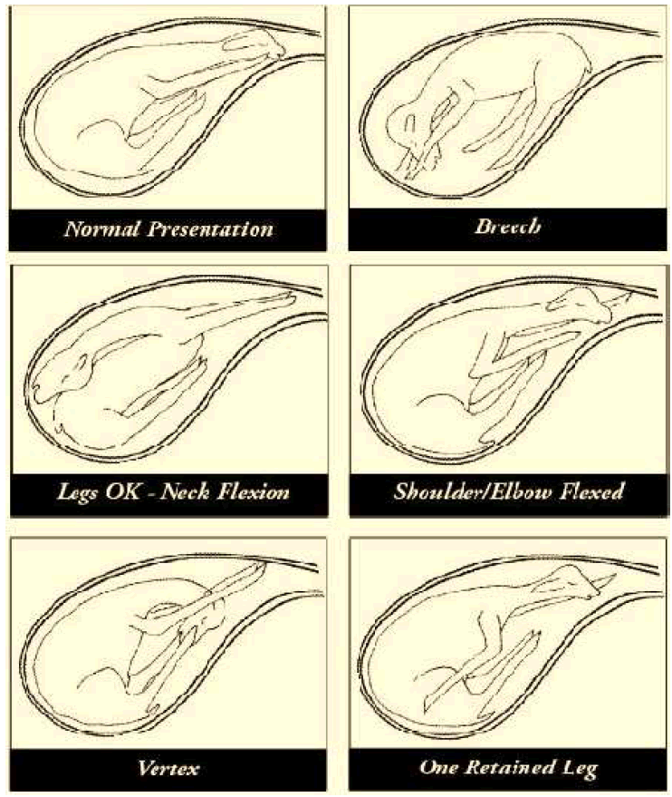
although the placenta also produces some progesterone. The normal birth presentation in uniparous animals is the
anterior longitudinal presentation, dorso-sacral position with the head resting on the metacarpal bones and knees of
the extended forelegs. Birth can occur without assistance if the fetus is in posterior longitudinal presentation dorsosacral
position and both hind limbs are extended. Unless, the fetus is small most other presentation, position and
postures result in dystocia. The transverse presentation can occur in the mare, in which the fetus develops in both
uterine horns, rather than in the body of uterus and one uterine horn. Transverse presentations are rare in ruminants,
and the small animals.
Following parturition the dam should be allowed to lick and nurse her young one. Undue excitement should be
avoided. Some animals have a strong maternal instinct and often object to shifting of their new born and this should
therefore be done slowly. The roughage fed should be of good quality.
Dystocias (Difficult births)
Dystocia may also be caused by maternal reproductive problems such as infection, poor nutrition or obesity where
excess fat in the birth canal reduces the area for the fetus to pass through.
Retained Placenta
The placental membranes are normally expelled within two to eight hours after birth. Occasionally, however, they
fail to separate from the uterus. If not treated, this condition may pose a health threat to the cow and cause problems
in rebreeding. The reason for retained placentas is not known, but high incidence may indicate a disease problem.
They also commonly accompany difficult births, multiple births, short gestations and bull calf births.
Research has shown that manual removal of retained placentas will decrease fertility. The recommended treatment is
to wait for about 48 hours after birth and then give injectable antibiotics along with uterine boluses or uterine
infusions. Observe the cow closely for swelling of the vulva or signs of illness.
Conclusion
Parturition is the most important event of an animal’s life. For livestock producers, it is a key event that can either
lead to economic gains, or to a loss should problems occur. By understanding how parturition occurs, it is easier for
livestock breeders to know when a problem occurs and what to do if an animal needs assistance. Although a large
emphasis in this paper is placed on giving assistance at birth, it is not to be implied that every animal will need
assistance with every birth. It is not uncommon for an animal to give birth to healthy offspring without any human
intervention.
References
- Asgari safdar, A.H., Daghigh Kia, H., Farhadi, R. Int J Adv Biol Biom Res. 2013, 1(3):214-221.
- Fuchs, AR. CRC Press, Boca Raton, Florida.1986, p. 163.
- Fuchs, AR, Fuchs, F. A review. Br J Obstet Gynaecol. 1984, 91:948.
- Garfield, RE, Kannan, MS, Daniel, EE. Am J Physiol. 1980, 238:C81.
- Huber, A, Hudelist, G, Czerwenka, K, Obstet Gynecol. 2005, 105:91.
- Jenkin G, Young IR. Anim Reprod Sci. 2004. 82 -85: 567- 81.
- Keirse, MJNC. Leiden University Press, Leiden, 1979, p. 101.
- Liggins, GC. N Engl J Med. 1993, 328:1509.
- MacLennan, AH, Nicolson, R, Green, RC. Equine Vet J Suppl. 1997, 83.
- Novy, MJ, Walsh, SW. Am J Obstet Gynecol. 1983, 145:920.
- Pitera, AE, Smith, GC, Wentworth, RA, Nathanielsz, PW. Am J Obstet Gynecol. 1998, 179:492.
- Potestio, FA, Zakar, T, Olson, DM. J Clin Endocrinol Metab. 1988, 67:1205.
- Roche JF. Anim Reprod Sci. 2006, 96: 282 -96.
- Spitz, IM, Bardin, CW. N Engl J Med. 1993, 329:404.
- Thorburn, GD, Challis, JRG, Robinson, JS. Plenum Press, New York. 1977, p. 653.
- Zeeman, GG, Khan-Dawood, FS, Dawood, MY. Obstet Gynecol. 1997, 89:873.


