Keywords
Annona muricata, Hepatoprotective, Stem bark, Acetamenophen, Anticholestasis, Antisinusoidalcongestion
Introduction
Migraine is a headache; severe enough to profoundly restrict or inhibit routine daily activity and with resultant nausea or light or sound sensitivity [1]. In the Global Burden of Disease Survey 2010, it was ranked as the third most prevalent disorder and seventh-highest specific cause of disability worldwide [2]. An analgesic is any medication intended for the relief of pain such as acetaminophen and opioid among others. Patients with chronic headache understandably tend to take analgesics frequently in an effort to reduce their pain and so enable themselves to carry out their routine activities [3].
Acetaminophen produces damage to the hepatic microvasculature (sinusoidal endothelial cell) which results in hepatocellular injury [4]. The reactive metabolite of acetaminophen, N-acetyl-p-benzoquinoneimine (NAPQI), is produced by cytochrome P-450 (CYP) as a result of direct two electron oxidation of acetaminophen [5]. The study carried out by Ito et al. [6] revealed swelling of the endothelial cells and erythrocytes penetration into the extrasinusoidal Space of Disse following acetaminophen overdose. More so, analgesic nephropathy, a form of chronic renal disease, has been reported to occur following recurrent liver injury due to excessive intake of analgesic mixtures [7]. Nephropathology, particularly proximal tubal cell necrosis, has been observed following acetaminophen overdose [5]. It is estimated that 9%, 3% and 1% of patients undergoing dialysis in Australia, Europe and the United States, respectively due to analgesic nephropathy [8]. Recent reports which favour the use of herbal preparations over synthetic drugs [9-11] have increased the number of researches carried out on plant materials in a bid to finding a safe natural analgesics and hepatoprotective products.
Annona muricata belonging to the family Annonaceae, found throughout the tropics, [12] and has been known to possess antidiabetic [12-14], antioxidant, antimutagenic [15], antinociceptic and antiulcerative effects [16,17]. It is usually recommended in cases of constipation, obesity, hypertension and coronary diseases [12]. However, little or no work has been carried out to evaluate the potency of aqueous stem bark extract of Annona muricata to ameliorate the toxicity caused by acetaminophen (paracetamol) consumption. Thus, the aim of this study is to determine the hepatoprotective effect of Annona muricata following acetaminophen induced hepatotoxicity.
Materials and Methods
Study area
The study was carried out at the animal facility in the department of Medical Laboratory Science, Faculty of Health Sciences, Madonna University Elele, Rivers state, Nigeria between May and July, 2015. The study area is located in the tropics (with the mean daily temperature of 29 ºC) at the southern part of Nigeria; latitude 5 27-5 31N and longitude 6 55-7 85E [18].
Ethical approval
The research received ethical approval from the departmental ethics committee of Madonna University, Nigeria. The experiment was conducted in accordance with the guidelines of the U.S. National Institute of Health [19] on the care and use of laboratory animals and also in accordance with the principles of Good Laboratory Procedure [20].
Animals handling
A total of 36 male albino rats, 2 to 3 weeks of age were used for this study. The experimental animals used for this study were obtained from the animal facility in Madonna University, Elele, Nigeria housed in wire meshed cage under standard conditions (temperature 25 -29°C, 12 hours light and 12 hours darkness cycles).
Test samples
The test samples were Acetaminophen and Annona muricata stem bark extract. The acetaminophen which is paracetamol was bought from Madonna University Pharmacy, was grinded and weighed. Fresh large quantity of stem bark of Annona muricata tree was gotten from Amucha village in Orlu local government of Imo state Nigeria. The botanic identification and authentification was carried out in faculty of pharmacy Madonna University, Elele campus by a taxonomist and a sample keep at the herbarium (MU/PHGSY/05/001).
Preparation of extract
Fresh stem bark of Annonamuricata was cut from the tree, dried for one week until there were no evidence of obvious moisture in them, the dried stem bark was grinded into powered form. Six hundred (600) ml of distilled water was added to 100 grams of the ground stem bark and allowed to stand in a refrigerator for 72hrs. The supernatant was decanted into a beaker, filtered using filter papers and the water evaporated to dryness in an oven. After drying, the powder form of the extract was wraped in aluminum foil and preserved in a refrigerator [21,22].
Experimental design
This experimental controlled study was carried out in Madonna University, Elele Campus, River State; A total of 36 male albino rats with an average weight of 88.03g were used as animal model, housed in six meshed cages containing 6 rats each. They were allowed to acclimatize for two weeks before the commencement of the experiment and fed on standard rat chow and water ad libitum for the duration of the experiment. Group 1, 2, 3, 4, 5 and 6 were orally administered 1ml of distilled water for 14 days, 300 mg/kg of acetaminophen for 7 days, 300 mg/kg of acetaminophen for 7 days and allow to heal naturally, 300 mg/kg of acetaminophen for 7 days and 25 mg/kg of Annona muricata extract for 7 days, 25kg body weight of Annona muricata extract for 7 days, 25mg/kg body weight of extract for 7 days and 300 mg/kg of acetaminophen for 7 days, respectively. The Annona muricata extract and acetaminophen were dissolved in 1 ml of distilled water before administration.
Sample collection and biochemical analyses
At the end of the experiment (14 days), the animals were subjected to an overnight fast, weighed using a weighing balance (Doran-PC 500), anesthetized by the “Drop method” [23], blood samples taken from their jugular vein and dispensed into plain containers labeled appropriately and allowed to coagulate. The samples were centrifuged in a Power Spin Centrifuge (C858E) for 10 minutes at 1500g within two hours after collection. The serum obtained was stored frozen until analyses. Immediately after the blood collection the rats were sacrificed and dissected. The liver was excised, blotted dry, weighed on a microbalance sensitive at 0.001 mg (Precisa 125A, Switzerland) and recorded. Liver samples were fixed in 10% formal saline until histopathological analysis. Biochemical analyses were carried out to determine the serum concentrations and activity of some enzymes such as AST and ALT (Randox Laboratories Ltd, United Kingdom, 303909) using the Reitman and Frankel method. ALP (Randox Laboratories Ltd, United Kingdom, 3230003) and LDH (Agappe Diagnostics, Switzerland, 51407002) level were estimated using the kinetic method as described by Sood [24]. Their absorbencies were read using a spectrophotometer (APEI PD-303S).
Automatic tissue processing
The tissues were processed in an automatic tissue processor (Jung Histokinette 2000) as follows: Beaker 1 (containing10% formal saline) for 3 hours, Beaker 2 (containing 70% alcohol)for1hour, Beaker 3 (containing80% alcohol) for 2 hours, Beaker 4 (containing90% alcohol) for2 hours, Beaker 5(containing95% alcohol) for2 hours, Beaker 6 (containing95% alcohol) for 2 hours, Beaker 7 (containingabsolute alcohol I) for 2hours, Beaker 8 (containing absolute alcohol II) for 2hours, Beaker 9 (containing xylene I) for 3 hours, Beaker 10 (containing xylene II) for 3 hours, Wax bath I for 3 hours, Wax bath II for 3 hours. After to the last processing stage, tissues were covered in molten paraffin wax in metallic embedding moulds, allowed to cool and solidify in a refrigerator for 15 minutes at 5ºC. The cooled and solidified tissue blocks were trimmed to remove excess wax and serially sectioned at 5 μm on a rotary microtome (KEDEE KD- 3668AM). The sections were floated in 20% alcohol and transferred to a water bath (HH-420) at 45ºC and picked up at an angle using clean frosted end slides. Adequate attachment of tissues to slides was ensured by placing the tissue and slide on a hot plate for a duration of 40 minutes.
Haematoxylin and Eosin staining
The tissue sections were dewaxed in 2 changes of xylene for 2 minutes each, hydrated through decreasing grades of alcohol (that is, 100%, 90%, 80% and 70%) for 2 minutes each, stained in Ehrlichshaematoxylin for 30minutes and rinsed in running tap-water to remove excess stain. The sections were differentiated in 1% acid alcohol for a second, blued in running tap water 10 minutes, counterstained with 1% eosin for 2 minutes, rinsed in water, dehydrated in ascending grades of alcohol (70%, 80, 95% and absolute), dealcoholized in xylene, dried at room temperature and mounted using dibuthylphthalate propylene xylene. The slides were examined under a light microscope and photomicrographs were taken using a camera attached to the microscope.
Statistical Analysis
The biochemical data were subjected to some statistical analysis: analysis of variance (ANOVA) and Post Hoc test was carried out on the data using the Statistical Package for Social Sciences (SPSS; version 18). Values were reported as Mean ± SD. P-value is significant at p<0.05.
Results
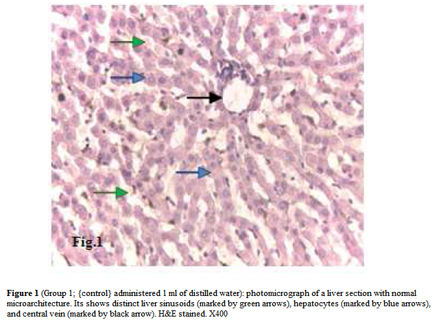
Figure 1: (Group 1; {control} administered 1 ml of distilled water): photomicrograph of a liver section with normal microarchitecture. Its shows distinct liver sinusoids (marked by green arrows), hepatocytes (marked by blue arrows), and central vein (marked by black arrow). H&E stained. X400
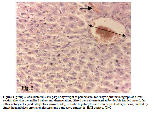
Figure 2: (group 2; administered 300 mg/kg body weight of paracetamol for 7days): photomicrograph of a liver section showing generalized ballooning degeneration; dilated central vein (marked by double headed arrow), few inflammatory cells (marked by black arrow heads), necrotic hepatocytes and iron deposits (karyorhesis; marked by single headed black arrow), cholestasis and congested sinusoids. H&E stained. X400
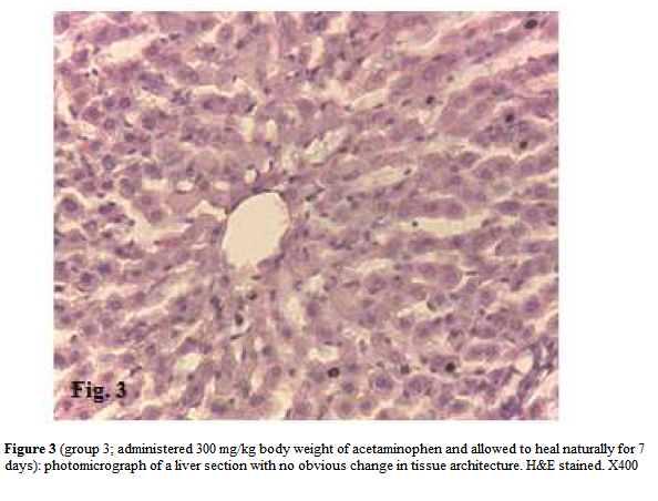
Figure 3: (group 3; administered 300 mg/kg body weight of acetaminophen and allowed to heal naturally for 7 days): photomicrograph of a liver section with no obvious change in tissue architecture. H&E stained. X400
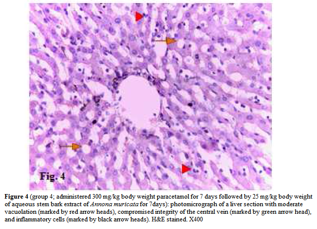
Figure 4: (group 4; administered 300 mg/kg body weight paracetamol for 7 days followed by 25 mg/kg body weight of aqueous stem bark extract of Annona muricata for 7days): photomicrograph of a liver section with moderate vacuolation (marked by red arrow heads), compromised integrity of the central vein (marked by green arrow head), and inflammatory cells (marked by black arrow heads). H&E stained. X400
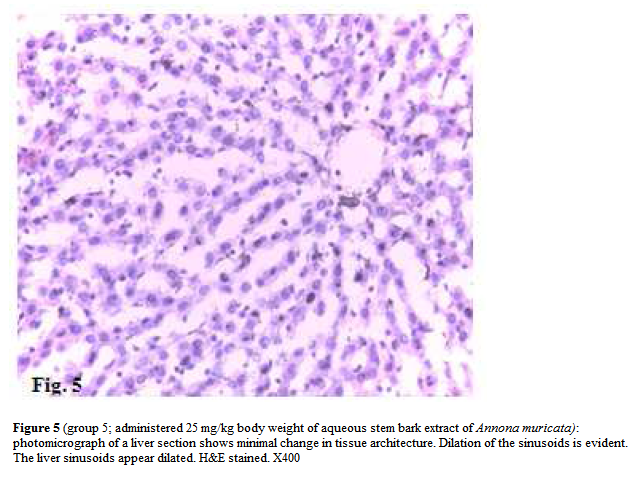
Figure 5: (group 5; administered 25 mg/kg body weight of aqueous stem bark extract of Annona muricata): photomicrograph of a liver section shows minimal change in tissue architecture. Dilation of the sinusoids is evident. The liver sinusoids appear dilated. H&E stained. X400
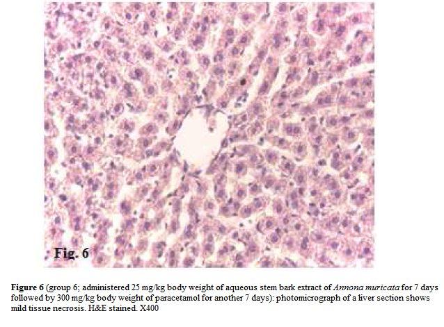
Figure 6: (group 6; administered 25 mg/kg body weight of aqueous stem bark extract of Annona muricata for 7 days followed by 300 mg/kg body weight of paracetamol for another 7 days): photomicrograph of a liver section shows mild tissue necrosis. H&E stained. X400
Discussion
Abuse of drug could range from small doses of paracetamol to large doses of morphine. These drugs may have minimal to severe effects on the liver and kidney following the formation of NAPQI. This study aimed to exploit the fact that Annonamuricata has been reported to possess antiulcer and antioxidant activity [16,17] to assess its potency in ameliorating the effect caused by acetaminophen (paracetamol) on the liver.
Hepatotoxicity following acetaminophen overdose or abuse in humans and rodents is associated with hepatic congestion. The congestion results from the accumulation of red blood cells within endocytic vacuoles and the Space of Disse with a collapse of the sinusoidal lumens [5]. The latter reports are in consonance with the findings of this study (figure 2) which showed decrease sinusoidal space with concomitant infiltration of red cells (an evidence of cholestasis) when compared with the control group (figure 1). The observed architectural change in the liver is also consistent with the drug induced hepatotoxicity described by Kumar et al. [25]. The study carried out by Walker and co-workers revealed that a very high dose of acetaminophen after one and half hours lead to an initial significant increase in liver weight and elevation of alanine transaminase and aspartate transaminase is associated with lysis of hepatocytes and necrosis [26,27]. The latter is consistent the significant increase in liver weight and serum enzyme levels observed in group 2 (Table and figure 2). The higher bodyweight and organ: body weight ratio also confirms that acetaminophen can be highly toxic in cases of overdose, prolonged use or drug-abuse.
Based on the result of this study (table and figure 5), it could be argued that aqueous stem bark extract of Annona muricata had no obvious pathological effect on the microarchitecture of the liver. As a matter of fact, it could also be suggested that the plant extract may have the potency of forestalling apoptosis or ameliorating hepatic damage, since the serum levels of ALP and ALT (key liver enzymes) were lower when compared with that of the control group. The reason for the higher serum level of AST and LDH activity following administration of the extract is still unclear. Aspartate aminotransferase (AST) and LDH are largely found in significant amounts in the heart and liver while lesser amounts are found in skeletal muscle, kidneys, pancreas, spleen, lungs and brain [28]. Furthermore, the administered dose of the extract increased the diameter of the sinusoids without significant change in tissue architecture. This latter effect of the extract could counter the cholestatic and sinusoidal congestion induced by acetaminophen on the liver.
The result of the study show that aqueous stem bark extract of Annona muricata may possess little or no hepatocurative potency at the administered dosage. This is due to the fact the group 4 had higher serum levels of ALP, AST and ALT compared to the animals that were allowed to heal naturally following artificially induced hepatotoxicity (group 3). Despite the lower activity of lactate dehydrogenase (group 4), microscopy of the liver section of group 3 showed no change in tissue architecture (figure 3) compared with that of group 4 that showed features of inflammation and drug toxicity (figure 4). The fact that the organ: body weight ratio of group 4 was observed to be higher compared with that of group 3 further undermines the hypothesis that Annona muricata may possess hepatocurative effect.
The biochemical analysis also revealed lower ALP, AST, ALT and LDH (table) in group 6 (administered 25 mg/kg body weight of aqueous stem bark extract of Annona muricata before artificially inducing hepatotoxicity) when compared with other groups that received paracetamol. This suggests that Annona muricata possess hepatoprotective effect at the administered dosage. This is supported by the liver microscopy of group 6 (figure 6) which showed less microarchitectural changes compared with group 2. In fact, there were significant differences between group 6 and group 2 in terms of tissue architecture, liver weight and enzyme level or activity (Figures 2 and 6, and table). Furthermore, there is no significant difference between group 1 (control) and group 6 in all the assessed parameters unlike when group 1 was compared with other groups. The observed anticholestatic and antisinusoidal congestion could be linked to the same phytochemical agents that confer antiulcer and antioxidant properties on Annona muricata. An example, is a class of bioactive agent known as alkaloid which include compound such as epoxymurin, reticuline and anomuricine among others [16,17, 28,29].
Furthermore, the increase in liver weight and elevation of liver enzymes following acetaminophen administration and reverse decrease observed when aqueous stem bark extract of Annona muricata was administered are in consonance with the report of Arthur et al., [30] who carried out their study using aqueous leaf extract of Annona muricata. This lends support to the work of Moghadamtousi et al. [29] who stated that alkaloids are found in both leaf and stem bark extracts of Annona muricata. The enzyme lactate dehydrogenase, can be found in almost all living cells, especially the liver, heart, red blood cells, brain, lungs, kidneys, pancreas and striated muscles [31]. It is released during tissue breakdown or some disease process, particularly in the case of hemolysis [32]. The decrease in transaminases and LDH observed in this study relates well with the decrease in liver enzymes, protein and malondialdehyde in both the liver and brain reported by Padma et al. [33] following ethanol stembark extract of Annona muricata administration. Thus, Annona muricata cannot be only used to forestall liver damage but also other organ damage following toxic chemical exposure. The decrease in LDH in this study also relates well with the reduction of jaundice as reported by Arthur et al. [34], after administration of aqueous extract of A. muricata leaf to rats suffering from artificially induced hepatic injury. This is due to the potency of A. muricata to reduce the rate of haemolysis (red blood cell breakdown). As This further supports the fact that all parts of A. muricata are medicinal [29].
Conclusion
The biochemical investigations and liver microscopy of this study suggests that aqueous stem bark extract of Annona muricata possess antihaemolytic, anticholestasis and antisinusoidal congestion properties. Thus, these properties of Annonamuricata extract can be exploited to treating several tissue breakdowns or side effects following drug administration.
Acknowledgements
Special thanks are to the management and staff of Medical Laboratory Services, Madonna University Teaching Hospital and Federal Medical Centre Owerri, Imo State, for allowing us make use of their facilities and also for their technical support.
References
- Rothrock, R.F. 2007b, 331. Doi: 10.1111/j.1526-4610.2007.01025.x
- International Headache Society. Cephalalgia. 2013, 33(9) 629–808. DOI: 10.1177/0333102413485658
- Rothrock, R.F. 2007a, 467-468 doi: 10.1111/j.1526-4610.2007.00748
- McCuskey, R.S. Clin Hemorheol Microcirc. 2006, 34(1-2):5-10. PMID: 16543612.
- Hinson, J.A., Roberts, D.W. and James, L.P. Handb Exp Pharmacol. 2010, (196): 369:405. Doi: 10.1007/978-3- 642-00663-0_12. PMCID: PMC2836803
- Ito, Y., Bethea, N.W., Abril, E.R. and McCuskey RS. Microcirc. 2003, 10:391–400. PMCID: 14557822
- De Broe ME and Elseveirs MM. N Eng J Med 1998, 338: 446
- Kumar K, Abbas AK, Fausto N. Robbins and Cotran Pathologic Basis of Disease. 7th ed. Saunders Elsevier, Philadelphia, PA, USA. 2005. 960
- Mangunwidjaja, S.D., Raharja, S., Kardono, L.B.S. and Iswantini, D. J. Applied Sci., 2006; 6: 1576-1580
- Omonkhua, A.A., Onoagbe, I.O. Internet J Nutr Wellness 2008; 6: 1937-8297.
- Upadhyay, H.C., Saini, D.C., Srivastava, S.K. Res J Phyochem, 2011, 5: 170-176.
- Pamplona-Roger, G.D. 2005. Available online at: www.amazon.com/encyclopedia-volume/dp/8472081842. Accessed on 21 August, 2015
- Shirwaikar, A., Rajendran. K and Dinesh KC. Pharmaceut Biol 2004, 42(1): 30-55
- Adewole, S.O., Caxton-Martins, E.A. and Ojewole, J.A. Pharmacol-online. 2006, 2: 1-21
- Thakkar, J.H., Solanki, H.K., Tripathi, P., Patel, N.J. and Jani, G.K. Elixir Human Physiol 2011; 31: 1960- 1965.
- Feng, P.C. J Pharmacol, 1999, 14(1): 556-561.
- Hamid RA, Foong CP, Ahmad Z, Hussain MK. Brazilian J Pharmacog. 2011, 22(3). Doi:10.1590/S0102- 695X2012005000001. www.scielo.br/scielo.php?pid
- Gobo, A.E. J Meterol. 1988, 13: 220-224.
- National Institute on Health: Guide for the care and use of laboratory animals (NIH publication No. 86-23). Bethesda, MD: Public Health Service 1985.
- WHO: Basic OECD Principles of GLP. Geneva, Switzerland: World Health Organization 1998. Available at: www.who.int/tdrold/publications/pdf/pr15/info.pdf (accessed 24 August, 2015)
- Francis, F.G. Cereal Food World. 2000; 45: 208-213.
- Cheng, C.W., Saad, S.M. and Abdullah, R. European J Biotechnol Biosci. 2014; 1(5): 14-18
- Johns Hopkins University (JHU). Animal care and use committee: Use of Anesthetic Gases: “Drop Method”. October 15, 2009. web.jhu.edu/animalcare/.../Anesthesia.Dr. (Accessed 15 May. 2015)
- Sood, R. Medical laboratory technology method and interpretations, 6th ed. New Delhi: V2, Jaypee Brothers Medical Publishers (P) Ltd, 2009.
- Kumar, K., Abbas, A.K., Fausto, N. And Aster, J.C. Robbins and Cotran pathologic basis of disease, 8th ed. Philadelphia, PA: Saunders Elsevier, 2010.
- Roberts, D.W., Bucci, T.J., Benson, R.W., Warbritton, A.R., McRae, T.A., Pumford, N.R. et al. Am J Pathol 1991, 138:359–371. PMCID: 1992763
- Gujral, J.S., Knight, T.R., Farhood, A., Bajt, M.L. and Jaeschke, H. Toxicol Sci 2002, 67: 322–328. PMCID: 12011492
- Wilkinson, J.H. Principle and practice for diagnosis enzymology. 2nd ed. Philadelphia. JB Lippincott Co. 1976; 282-319.
- Moghadamtousi, S.Z., Fadaeinasab, M., Nikzad S., Mohan, G., Ali, H.M., Kadir, H.A. Int Mol Sci, 2015; 16(7): 15625-15658.
- Arthur, F.K.N., Woode, E., Terlabi, E.O., Larbie, C. J Nat Pharm 2012a; 3: 25-30.
- Van Eerd, J.P., Kreutzer, E.K. Klinische Chemie voor Analisten deel 2. 1996; 138-139.
- Kato, G.J., McGowan, V., Machado, R.F., Little, J.A., Taylor, J., Morris, C.R., et al. Lactate dehydrogenase as a biomarker of haemolysis. Blood 2006; 107(6): 2279-2285.
- Padma, P.; Chansouriab, J.P.N., Khosa, R.L. Anc Sci Life 1999; 19(1-2): 7-10. PMC3336466
- Authur F.K.N., Woode, E., Terlabi, E.O., Larbie, C. Am J Pharm Toxicol 2012b; 7 (2): 33-40

