 |
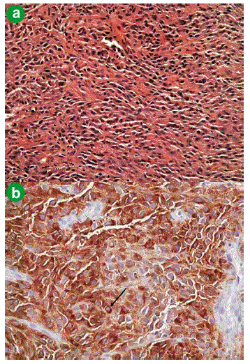
| Figure 3. Pathology slides confirming the final diagnosis of malignant melanoma including immunohistochemistry stain for S- 100. a. The tumor is composed of pleomorphic, loosely cohesive cells with enlarged, hyperchromatic, irregular nuclei (20x, Hematoxylin and Eosin). b. Tumor cells show positive staining for Melan-A protein. Arrow depicts cytoplasmic staining (20x, immunohistochemistry). |