|
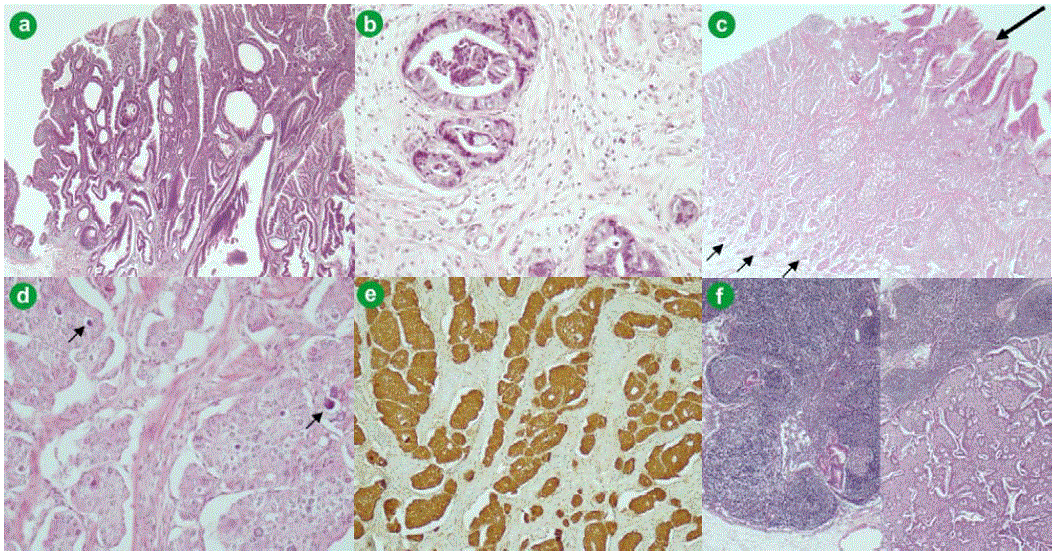
| Figure 2.a. The high grade tubulo-villous adenoma extending to the duodenal mucosa (H&E staining, x200). b. An area of well differentiated adenocarcinoma infiltrating the duodenal submucosa (H&E, x400). c. The endocrine component (small arrows) is located under the exocrine high grade adenoma (large arrow) (H&E, x100). d. The endocrine component is well differentiated and contains psammoma bodies (arrows) (H&E, x400). e. Endocrine tumor cells abundantly and diffusely expressing somatostatin (immunostaining with somatostatin antibody, counterstained with hematoxylin; x 200). f. Metastatic lymph nodes are present, either from the exocrine (left) or the endocrine (right) component (H&E, x100). |