 |
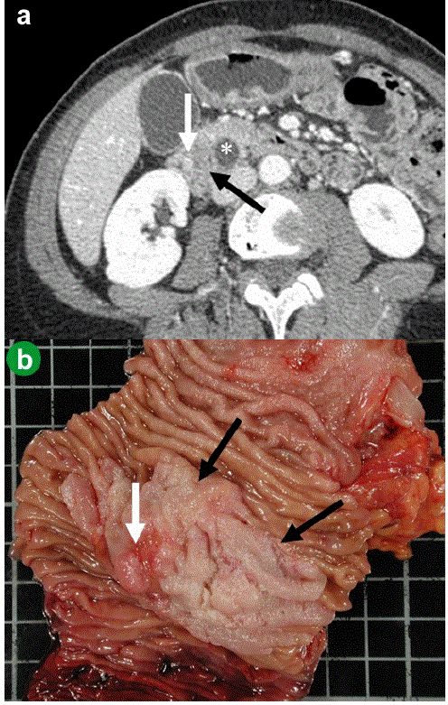
| Figure 1. a. CT-scan slice at the level of the ampulla, obtained during the arterial phase after an intravenous injection of contrast medium, identified a large ampullary and peri-ampullary tumor invading the duodenum (large black arrow). This tumor is heterogeneous with a distinct hyperdense nodule (white arrow). The common bile duct (*) is dilated. b. The large exophytic duodenal tumor is situated in and around the ampulla of Vater, measuring 4.5x5.0 centimeters. At the center of this tumor, in close contact with the ampulla of Vater, a well delineated red-brown nodule measuring 12 mm and corresponding to the endocrine component is visible. |