 |
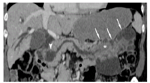
| Figure 1. Curved coronal CT image of the pancreas in the portal venous phase of contrast enhancement. Communication of the mainly cystic intraductal oncocytic papillary neoplasm to the main pancreatic duct in the pancreatic head (arrowhead) and tail is clearly depicted (arrows). Please note the considerable size of the solid tumor part in the tail of the pancreas (asterisk). |