 |
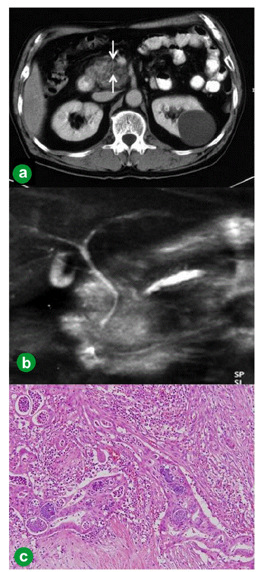
| Figure 1. Case#1: initial operation. a. A tumor approximately 1 cm in diameter was detected in the pancreatic head on abdominal CT (arrow). b. The main pancreatic duct was disrupted, and the main pancreatic duct of the distal pancreas was dilated on MRCP. c. Welldifferentiated tubular carcinoma. Marked fibrosis and acinus atrophy were observed. Mild atypical cells with a slightly swollen nucleus formed an irregular ductal structure and invaded the parenchyma. (H&E stain, x100). |