|
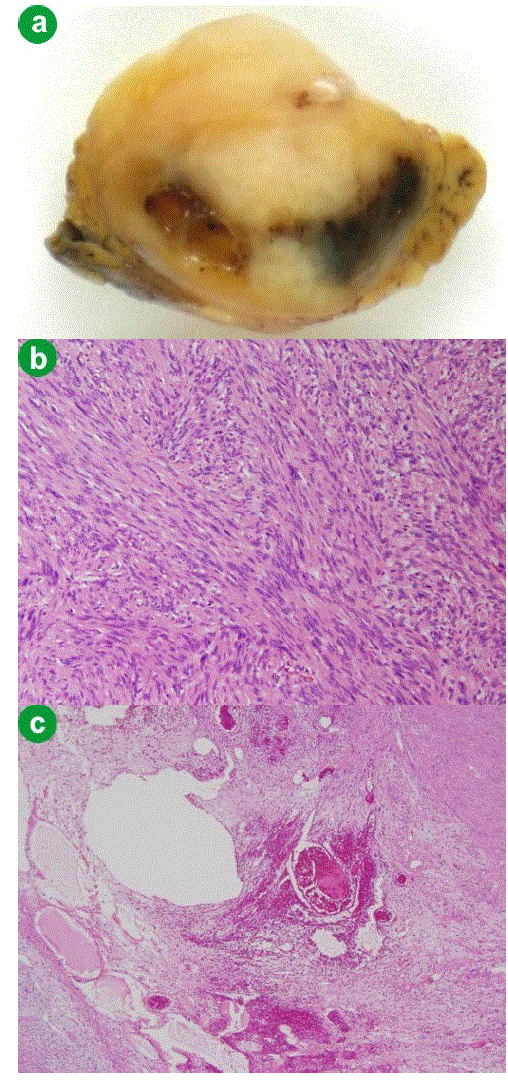
| Figure 3a.a. The cut surface of the resected tumor is composed of a mixture of solid areas and myxomatous and/or hemorrhagic areas. b. Histopathology of the resected specimen shows proliferation of the spindle cells in interlacing and palisading patterns (Antoni A) (H&E, original magnification x100). c. Edematous degeneration areas with hemorrhage, hemosiderin deposition and hyalinization of the dilated vascular walls (Antoni B) (H&E, original magnification x100). |