 |
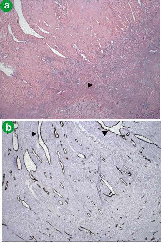
| Figure 2. a. H&E staining of the residual tumor in the region of head of pancreas/duodenum. In the lower right portion of the figure there is dense fibrosis due to treatment effect (arrow). Residual tumor is difficult to appreciate. b. A cytokeratin AE 1/3 immunohistochemical stain (at 40x) of the same region. One can see that the slit-like spaces seen on H&E stain in the muscularis propria of the duodenum are lined by residual tumor cells (arrow). |