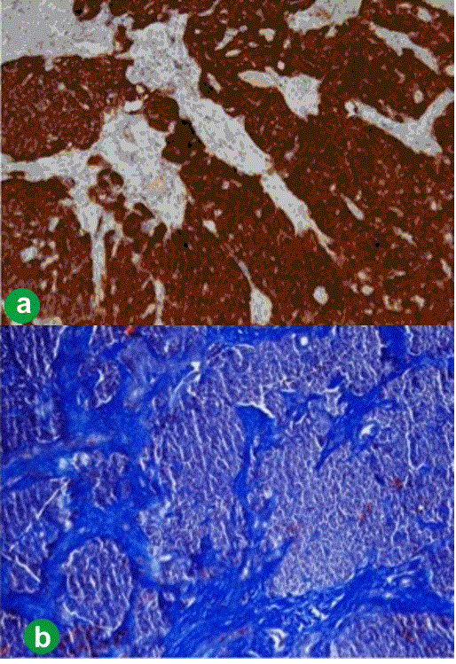
Figure 5.
Microscopic section of the tumor. a. Immunohistochemical coloring with anti-CD56 antibodies showing the presence of neuroendocrine tumor cells. b. Blue stain, Masson trichrome, showing dense fibrous strands.