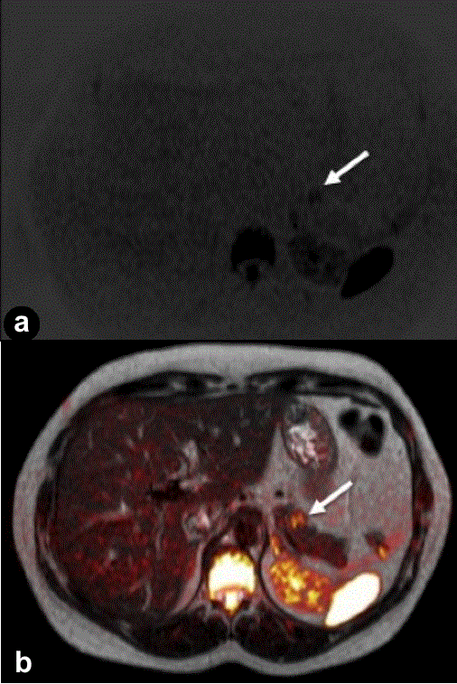
Figure 3.
Axial diffusion-weighted imaging (DWI) (a.) and fused image (b.) clearly show the tumor (arrows) with marked high signal intensity in the pancreatic tail.