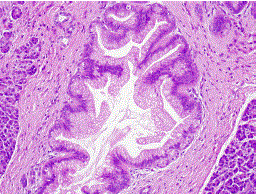
Figure 3.
Histology of intraductal papillary mucinous neoplasm showing low grade dysplasia (H&E stain, magnification x200; Patient #1).