 |
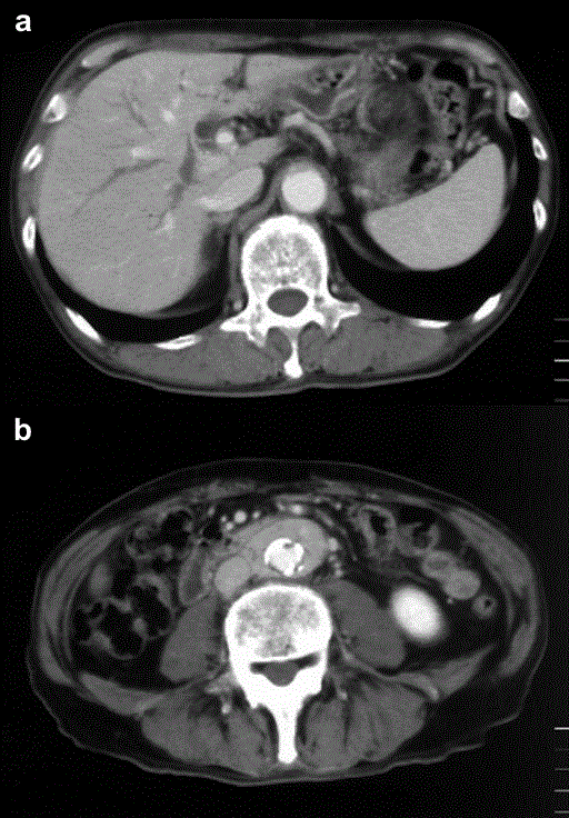
| Figure 1. Abdominal enhanced CT imaging with contrast medium in October 2003. a. Intra-hepatic bile duct dilatations were seen; however, the lesion responsible which impaired biliary flow was not identified. A Soft tissue mass having good enhancement surrounding the abdominal aorta and common iliac artery was incidentally demonstrated. b. There were no signs of hydronephrosis suggesting encasement of the ureter. |