|
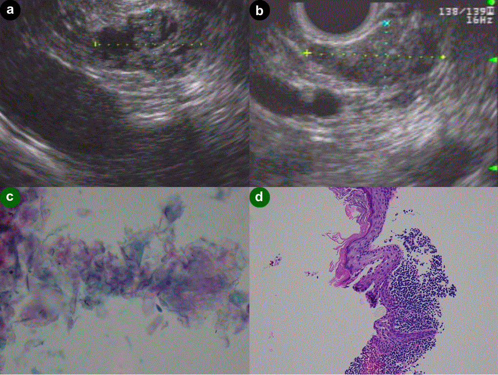
| Figure 1. Case 1. a. EUS: pancreatic tail lesion. 2.3x1.5 cm hypoechoic, slightly lobulated contour, granular echotexture, cystic lesion. b. EUS: pancreatic body lesion. Hypoechoic lesion with a granular echotexture. 2.0x1.0 cm in size. c. On-site touch imprint cytology: anucleate squamous cells. d. Histology: stratified squamous epithelium with prominent keratinaceous debris and adjacent lymphoid infiltrate without atypia. |