|
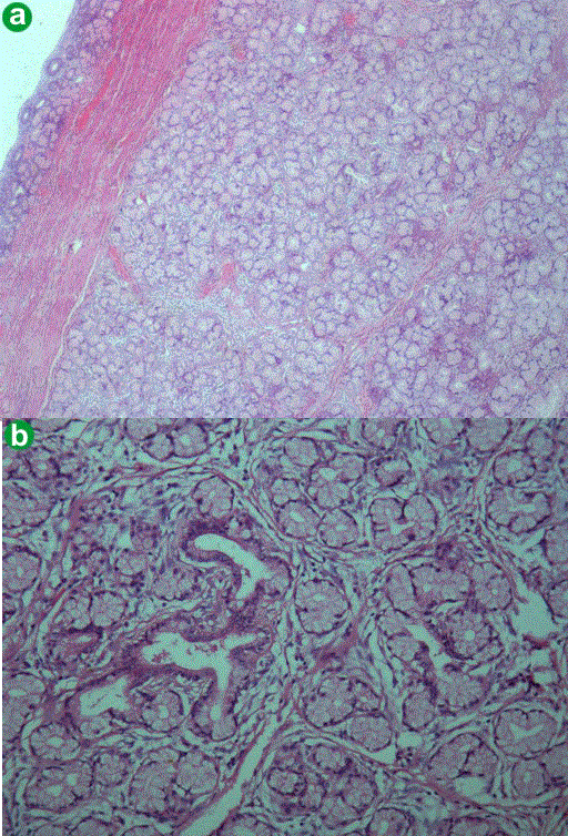
| Figure 4. a. Low power photomicrograph to show stretched-out duodenal mucosa overlying the mass lesion (H&E, x60). b. High power photomicrograph to show lobular collection of normal looking mucous glands with a draining intralobular duct (H&E, x240). |