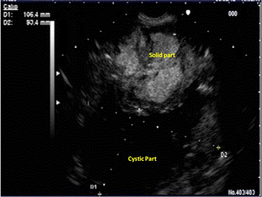
Figure 2.
Curved linear array endosonographic image of a complex heterogeneous mass of the retroperitoneum with hyperechoic and cystic component (9x10 cm).