|
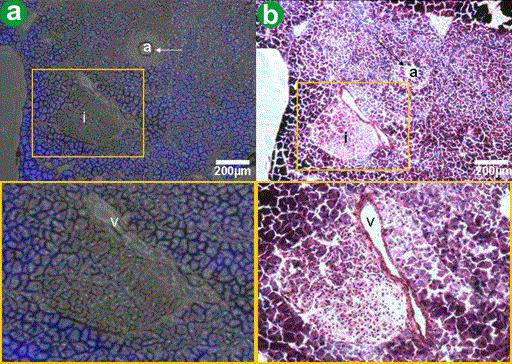
| Figure 7. H&E staining of the pancreas. H&E staining of the pancreas was performed to identify which components were responsible for the cyan fluorescence. a. The image shows the microscopic fluorescence of pancreatic tissue. The adjoining picture is a blow-up of the inset box and focuses on an area of nonfluorescence. b. The H&E stain of this exact same area of the tissue demonstrates that the cyan fluorescence is due to the pancreas’ exocrine parenchyma (acinar cells), and not from islet cells (i), seen in the blow-up box of the image. The vein (v), collapsed in this picture, and an artery (a) do not display cyan fluorescence either. Although not shown in this figure, the pancreatic duct cells did not display cyan fluorescence. |