|
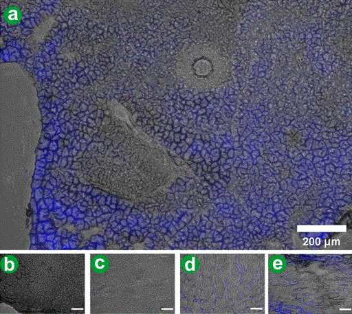
| Figure 6. Fluorescence microscopy of selected tissue. Selected organs were processed for fluorescence microscopy with an IMT-2 (Olympus Corp., Tokyo, Japan) inverted fluorescence microscope. The microscopic images were obtained in parallel with whole organ images, clearly demonstrating cyan fluorescence in the pancreas (a.). By contrast, microscopic images of the liver (b.) and spleen (c.) failed to pick up any blue signal. The muscle (d.) and peritoneum (e.) displayed cyan fluorescence. |