 |
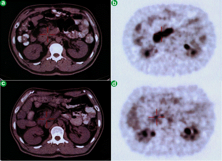
| Figure 7. a. A CT scan made before high intensity focused ultrasound demonstrating a tumor in the head of the pancreas. b. A PET-CT scan made before high intensity focused ultrasound demonstrating a SUVmax of 9.1 g/mL. c. A CT scan demonstrating no significant change one month following high intensity focused ultrasound treatment. d. The PET-CT scan made one month after high intensity focused ultrasound demonstrated that the SUVmax value decreased to 3.1 g/mL. All four images are taken from the same patient. |