|
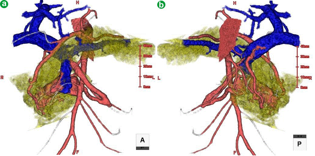
| Figure 3. Volume rendering models. a. Image taken from an anterior view. b. Image taken from a posterior view. The arterial system, portal venous system and pancreatic parenchyma were integrated into the model. Note the clear visualization of the spatial relationship between the aberrant vascular network and the pancreatic parenchyma. |