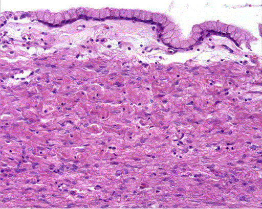
Figure 5.
Computed tomography scans (a. axial; b. coronal multiplanar) reveal an inhomogeneus thickening of the duodenal wall by multiple cystic lesions compressing the duodenal lumen, common bile duct and Wirsung duct.