|
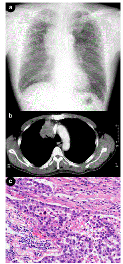
| Figure 1. a. Chest roentgenography showed an irregular bulging mass at the right hilum of the lung, next to the superior vena cava. b. A chest CT-scan identified a mass displacing the superior vena cava, suspicious of superior vena cava invasion. c. Microscopic findings of the lung tumor, showing poorly differentiated adenocarcinoma with vascular infiltration (hematoxylin and eosin staining). |